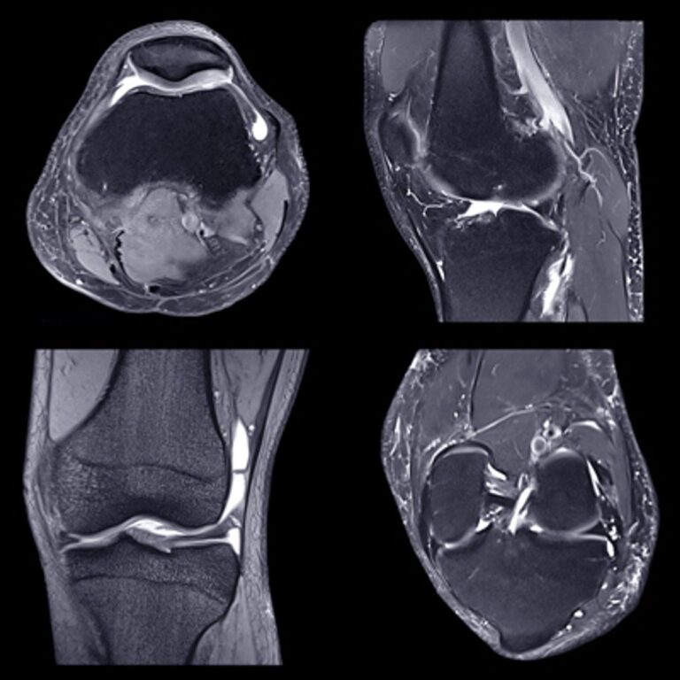
NYU Langone Advanced Imaging of the Musculoskeletal System Up Your Game 2023 is a comprehensive and intensive series of online CME lectures covering major topics, including common sports injuries, bone tumors, arthritis and cartilage imaging, pediatric imaging, and AI. You can explore new advances in MR, CT, and US, and update skills with continuing medical education that will help you to:
Format : 59 Videos + 2 PDFs
Description
videos + pdf, size: GB
+ Target Audience: radiologist, musculoskeletal radiology physician
Musculoskeletal Imaging CME: State-of-the-Art Updates
You can explore new advances in MR, CT, and US, and update skills with continuing medical education that will help you to:
- Improve therapeutic decision making by learning how to modify imaging techniques and protocols for all areas of musculoskeletal MRI
- Develop a working differential for bone and soft tissue tumors and know when additional imaging or tissue sampling is warranted
- Evaluate the use of imaging in diagnosis and treatment of inflammatory arthritides and common musculoskeletal infections
- Describe techniques that improve MR imaging in the presence of hardware, as well as areas of pathology that can be diagnosed post hip, knee, or shoulder arthroplasty
Date of Original Release: July 15, 2023
Estimated Time to Complete: 21.25 hours
Learning Objectives
After participating in this activity, clinicians should be able to:
- Determine how best to modify imaging techniques and protocols for all areas of Musculoskeletal MRI including: hip, knee, shoulder and foot, for improved therapeutic decision making
- Develop a working differential for tumors of both the bone and soft tissues and recognize when additional imaging or tissue sampling is warranted
- Evaluate the value of imaging in guiding diagnosis and treatment of such inflammatory arthritides as the rheumatoid and the spondyloarthropathies as well as common musculoskeletal infections, including, but not limited to the septic hip and discitis
- Describe techniques which can be utilized to improve MR imaging in the presence of hardware as well as the major areas of pathology which can be diagnosed in patients status post hip, knee and shoulder arthroplasty
Intended Audience
Radiologists, both in academic and private practice, seeking to increase their skills in musculoskeletal radiology, as well as orthopedists and physical medicine and rehabilitation practitioners interested in learning how imaging can best be incorporated into their practice.
Topics:
Shoulder
-
- MR of the Rotator Cuff – Pearls and Pitfalls – Michael J. Tuite, MD, FACR
- Tendon Delamination with Emphasis on the Rotator Cuff – Donald Resnick, MD
- The Rotator Cuff Interval – Anatomy and Pathology – Miriam A. Bredella, MD, MBA
- Imaging the Shoulder Labrum – Above the Equator – Donald Resnick, MD
- Anterior Shoulder Instability – Most Important Bone and Soft Tissue Injuries – Soterios Gyftopoulos, MD, MBA, MSc
- Post Operative Shoulder – How to Image and What Do I Need to Know? – Lawrence White, MD
Elbow, Wrist, and Hand
-
- Elbow Imaging in the Throwing Athlete – David A. Rubin, MD
- Ultrasound of the Elbow – Ronald S. Adler, MD, PhD
- Ulnar Sided Wrist Injuries – David A. Rubin, MD
- Fractures of the Wrist – What You Need to Know – Dana Lin, MD
- MRI and US of Commonly Encountered Finger Pathology – Catherine N. Petchprapa, MD
- Pediatric Sports Injuries of the Upper Extremity – Erin F. Alaia, MD
Hip
-
- Femoro-acetabular Impingement – What is the Current Thought, and How Should We Assess it – Lawrence White, MD
- Tendons of the Pelvis and Hip – Anatomy and Pathology – Miriam A. Bredella, MD, MBA
- Post Op Imaging of the Hip Arthroplasty – What the Surgeon Wants to Know – Leon D. Rybak, MD
- Groin Injuries in Athletes – Soterios Gyftopoulos, MD, MBA, MSc
- Bone Abnormalities of the Hip – David A. Rubin, MD
- Extra-articular Impingement Syndromes – Mohammad M. Samim, MD
AI and Metal Imaging
-
- AI Uses in MSK Imaging – Acceleration, Quantification, and Miscellaneous – Michael P. Recht, MD
- AI Pattern Recognition in MSK – State of the Art – Dana Lin, MD
- Metal Imaging on CT and MR – Iman Khodarahmi, MD, PhD
Knee
-
- Patterns of Knee Injury – Donald Resnick, MD
- Anterior Knee Pain and Patellofemoral Maltracking – Mini Pathria, MD
- MRI of the Knee – How Important are the Corners? – Lawrence M. White MD, FRCPC
- Menisci and Cruciates – Is There Anything New I Need to Know? – Christine B. Chung, MD
- Imaging of Cartilage and Cartilage Repair – What Does My Surgeon Need to Know? – Richard Kijowski, MD
- Cysts, Bubbles and Bursa – the Spaces Around the Knee – Mark D. Murphey, MD
Ankle and Foot
-
- Lateral Ankle Instability – the Highs and Lows – Lawrence White, MD
- Osteochondral Injuries of the Ankle and Foot – Donald Resnick, MD
- Imaging Heel Pain – Mark D. Murphey, MD
- Imaging of the Midfoot and Forefoot – Christine B. Chung, MD
- Common Pathology of Ankle Tendons – Michael J. Tuite, MD, FACR
Tumors
-
- Bone Tumors – When Should I Worry? – Mark D. Murphey, MD
- Soft Tissue Tumors – Pitfalls and Mimics – Mark D. Murphey, MD
- Tumor Biopsy – Dilemmas and Pitfalls – Christopher J. Burke, MD
- Image Guided Ablation of Bone Tumors – Mohammad M. Samim, MD
Scroll with the Experts
-
- Scroll with the Experts – Knee – Christine B. Chung, MD
- Scroll with the Experts – Shoulder – Miriam A. Bredella, MD, MBA
- MR of the Pelvis and Hip – Focus on Pelvic Tendons – Mini Pathria, MD
- Scroll with the Experts – Elbow – Michael J. Tuite, MD, FACR
- Scrolling Session – Ankle and Foot – Jan Fritz, MD
Imaging of Muscles and Tendons
-
- Muscle Injuries – Grading and Return-to-Play – Jan Fritz, MD
- Common Muscle Injuries of the Lower Extremity – Iman Khodarahmi, MD, PhD
- Common Muscle Injuries of the Upper Extremity – Michael J. Tuite, MD, FACR
- Pectoralis Injuries – Mini Pathria, MD
- Tendons Turning Tightly – William Palmer, MD
- Imaging of Nerves – Meghan Jardon, MD
Spine
-
- Cervical Spine Trauma – A Pattern Approach – Mini Pathria, MD
- Thoraco-Lumbar Spine Trauma – A Pattern Approach – Gina A. Ciavarra, MD, MMCi
- Spine MR Symptom-MRI Correlation – William Palmer, MD
- Discitis – Imaging Diagnosis and When to Biopsy – Christopher J. Burke, MD
- Sacroiliac Joint, Sacrum, and Coccyx – William R. Walter, MD
- What the MSK Radiologist Needs to Know About the Thecal Sac and Spinal Cord – Girish M. Fatterpekar, MD
Imaging of Arthritis and Infection
-
- Diagnosing Arthritis – My Approach – David A. Rubin, MD
- Imaging of Spondyloarthropathies – What’s New? – Christine B. Chung, MD
- Imaging in Rheumatology – A Radiologist Perspective – William Palmer, MD
- Imaging of Crystal Arthropathy – Gregory Chang, MD
- Myositis and Necrotizing Fasciitis – What Do I Need to Know? – William Palmer, MD
- The Diabetic Foot – Renata La Rocca Vieira, MD


