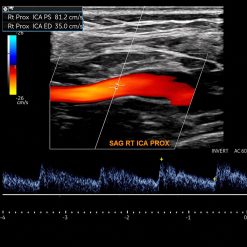

Advanced Sonography of the Posterior Shoulder and Scapula (Videos)


Original price was: $49.00.$15.00Current price is: $15.00.
This Product is shared via google drive download link, So please share your correct Gmail id while placing the order .Please note that there are no CME points or certificate associated with this course Samples for Courses Can be found here : Free Samples Here!
Advanced Sonography of the Posterior Shoulder and Scapula (Videos)
Advanced Sonography of the Posterior Shoulder and Scapula (Videos) is a specialized musculoskeletal ultrasound educational webinar presented by the American Institute of Ultrasound in Medicine (AIUM) in 2024. Featuring Phillip Steele, MD, RMSK, this presentation explores advanced posterior shoulder sonography, scapular anatomy, muscular insertions associated with the infraspinatus aponeurosis, and the recognition of normal and pathological ultrasound findings. The educational recording provides valuable insights into anatomical relationships, diagnostic imaging techniques, and clinical considerations relevant to shoulder pain, scapular dysfunction, and musculoskeletal assessment.
Target Audience
Musculoskeletal Radiologists, Diagnostic Sonographers, Sports Medicine Physicians, Orthopedic Surgeons, Physical Medicine and Rehabilitation (PM&R) Specialists, Rheumatologists, Pain Management Physicians, Primary Care Physicians, Physiotherapists, Musculoskeletal Ultrasound Specialists, Orthopedic Residents and Fellows, and healthcare professionals involved in shoulder imaging, scapular assessment, and musculoskeletal diagnosis.
Course Information
- Course Title: Advanced Sonography of the Posterior Shoulder and Scapula
- Organizer: American Institute of Ultrasound in Medicine (AIUM)
- Faculty: Phillip Steele, MD, RMSK
- Publication Year: 2024
- Format: High-Definition MP4 Video
- Included Files: 1 MP4 Video
- File Size: Approximately 1.9 GB (1948 MB)
- Language: English
- Specialty: Musculoskeletal Ultrasound / Shoulder Imaging / Orthopedic Sonography
The AIUM Advanced Sonography of the Posterior Shoulder and Scapula 2024 webinar provides a focused educational review of the complex anatomy and sonographic evaluation of the posterior shoulder and scapular region, emphasizing structures that contribute to shoulder stability, mobility, and coordinated muscular function.
The posterior shoulder contains interconnected muscular, tendinous, and fascial structures that play important roles in upper-extremity movement. Accurate ultrasound assessment requires detailed knowledge of these anatomical relationships and the ability to distinguish normal tissue appearances from findings that may suggest musculoskeletal pathology.
Presented by Phillip Steele, MD, RMSK, this educational activity places particular emphasis on the anatomy and physiology of posterior muscular insertions associated with the infraspinatus aponeurosis. Understanding these structures and their functional relationships provides an important foundation for interpreting posterior shoulder ultrasound images and evaluating regional musculoskeletal abnormalities.
The webinar examines relevant scapular anatomical landmarks, posterior shoulder muscular structures, and sonographic imaging characteristics, highlighting how careful anatomical identification contributes to effective diagnostic assessment. It also addresses the recognition of normal and pathological findings encountered during examination of the posterior shoulder region.
Another important focus is the role of diagnostic musculoskeletal ultrasound in evaluating posterior shoulder pain, restricted mobility, and scapular dysfunction. Sonographic examination can complement clinical assessment by providing real-time visualization of accessible soft-tissue structures and supporting further investigation when clinically indicated.
The educational content reinforces systematic ultrasound examination principles, accurate anatomical orientation, and careful image interpretation. These skills are especially valuable when assessing complex muscular and fascial relationships in patients with orthopedic or sports-related shoulder complaints.
Through expert-led instruction, this webinar supports radiologists, sonographers, orthopedic specialists, sports medicine clinicians, and rehabilitation professionals seeking to strengthen their understanding of advanced shoulder sonography, infraspinatus-associated anatomy, and diagnostic evaluation of the posterior scapular region.
Learning Objectives
Upon reviewing this educational activity, participants should be better able to:
- Identify important anatomical landmarks and musculoskeletal structures of the posterior shoulder and scapular region.
- Understand the anatomy and physiology of posterior muscular insertions associated with the infraspinatus aponeurosis.
- Recognize normal sonographic appearances of muscular, tendinous, and fascial structures in the posterior shoulder.
- Differentiate expected anatomical findings from potentially pathological sonographic abnormalities.
- Review systematic ultrasound examination principles relevant to posterior shoulder and scapular imaging.
- Understand the diagnostic role of musculoskeletal ultrasound in evaluating shoulder pain, restricted mobility, and scapular dysfunction.
- Relate anatomical relationships and sonographic findings to the clinical assessment of posterior shoulder disorders.
Key Topics Covered
- Advanced Posterior Shoulder Sonography
- Musculoskeletal Ultrasound Examination
- Posterior Shoulder Anatomical Landmarks
- Scapular Anatomy and Functional Relationships
- Infraspinatus Aponeurosis Anatomy
- Posterior Muscular Insertions and Fascial Structures
- Muscular and Tendinous Ultrasound Appearances
- Normal and Pathological Sonographic Findings
- Scapular Movement and Shoulder Function
- Ultrasound Identification of Posterior Shoulder Structures
- Systematic Shoulder Ultrasound Assessment
- Diagnostic Ultrasound Image Interpretation
- Posterior Shoulder Pain Evaluation
- Scapular Dysfunction and Musculoskeletal Assessment
- Orthopedic and Sports Medicine Ultrasound
- Clinical Correlation of Sonographic Findings
Clinical & Educational Value
This AIUM educational webinar provides focused instruction on the anatomical and sonographic assessment of the posterior shoulder and scapula, particularly the muscular insertions associated with the infraspinatus aponeurosis.
By emphasizing detailed anatomical knowledge, ultrasound identification of musculoskeletal structures, and recognition of pathological findings, the presentation supports a more systematic approach to shoulder imaging and clinical evaluation.
The educational recording is particularly relevant to musculoskeletal radiologists, diagnostic sonographers, sports medicine physicians, orthopedic specialists, and rehabilitation clinicians involved in assessing posterior shoulder pain, restricted movement, and scapular dysfunction.
Short Description
AIUM Advanced Sonography of the Posterior Shoulder and Scapula (Videos) 2024 is a specialized musculoskeletal ultrasound webinar presented by Phillip Steele, MD, RMSK. This 1.9 GB HD MP4 educational recording covers posterior shoulder anatomy, scapular sonography, infraspinatus aponeurosis, muscular insertions, normal and pathological ultrasound findings, and diagnostic assessment of shoulder pain and dysfunction. Designed for radiologists, sonographers, orthopedic specialists, sports medicine physicians, and rehabilitation professionals.
Related products


Anesthesiology & pain medicine
Gulfcoast Ultrasound-Guided Regional Anesthesia : Upper Extremities

Hematology / Oncology

Critical Care - Emergency medicine
The Brigham Board Review in Infectious Diseases Brigham and Women’s Hospital Board Review
Critical Care - Emergency medicine
2020 Radiology After Five: How to Make Night and Weekend Call a Success!

Critical Care - Emergency medicine