
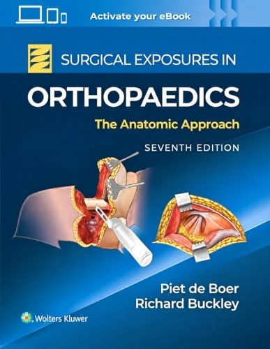
Surgical Exposures in Orthopaedics: The Anatomic Approach, 7th edition (Videos Only)
Original price was: $277.80.$50.00Current price is: $50.00.
Orthopaedic Surgical Exposures Videos, 7th Edition, demonstrate operative approaches, applied anatomy and structures at risk.
Delivery Information: This Product is shared via google drive download link. Please use your correct Gmail address at checkout.
Please note: there are no CME points or certificate associated with this course.
Samples for Courses Can be found here: Free Samples Here!
MedicalAmboss File Scope: This listing describes the educational subject matter for reference. Provider registration, live attendance, streaming-platform access, CME or other credit, certificates, memberships, apps, accounts, and publisher-hosted extras are not included unless they are explicitly listed as store files. MedicalAmboss.
Orthopaedic Surgical Exposures Videos 7th Edition
Surgical Exposures in Orthopaedics Videos | Orthopaedic Surgical Approaches | Applied Surgical Anatomy | Arthroscopic Approaches | Lower-Limb Amputation | Orthopedic Surgery
Information
Surgical Exposures in Orthopaedics: The Anatomic Approach, 7th Edition (Videos Only) provides practical visual instruction in the surgical anatomy and operative approaches used throughout orthopaedic surgery.
For more than four decades, Surgical Exposures in Orthopaedics: The Anatomic Approach has served as a major reference for understanding how surgeons safely reach bones, joints, and other musculoskeletal structures while protecting important nerves, vessels, muscles, and surrounding tissues.
The fully revised seventh edition continues the legacy established under Stanley Hoppenfeld, with Richard Eric Buckley and Peter Goffe de Boer (Piet de Boer) presenting orthopaedic anatomy specifically from the surgeon’s perspective.
The associated procedural videos provide visual demonstrations of surgical exposures from multiple angles, helping learners better understand incisions, anatomical landmarks, internervous and intermuscular planes, superficial and deep dissection, and structures at risk.
Product Details
- Title: Surgical Exposures in Orthopaedics: The Anatomic Approach
- Edition: 7th Edition
- Format Available Here: Videos Only
- Authors/Editors: Richard Eric Buckley, Peter Goffe de Boer
- Legacy Author: Stanley Hoppenfeld
- Publisher: Lippincott Williams & Wilkins / Wolters Kluwer
- Publication Year: 2026
- Specialty: Orthopaedic Surgery / Surgical Anatomy
- Full Book Length: 896 pages
- Language: English
Wolters Kluwer lists the seventh edition as a 2026 publication and categorizes it within orthopaedics, anatomy, residents, and surgery.
Target Audience
This procedural video collection is particularly relevant for:
- Orthopaedic Surgeons
- Orthopaedic Trauma Surgeons
- Sports Medicine Surgeons
- Joint Reconstruction Surgeons
- Foot and Ankle Surgeons
- Shoulder and Elbow Surgeons
- Hand and Upper Extremity Surgeons
- Pediatric Orthopaedic Surgeons
- Spine Surgeons
- Musculoskeletal Oncology Surgeons
- Orthopaedic Residents
- Orthopaedic Fellows
- Surgical Trainees
- Clinicians studying operative musculoskeletal anatomy
Program Overview
Safe orthopaedic surgery depends on more than knowing where an incision should be made.
Surgeons must understand:
- Surface anatomy
- Deep anatomy
- Muscular intervals
- Internervous planes
- Neurovascular structures
- Surgical landmarks
- Structures at risk
- Proper direction of dissection
- Safe retraction
- Extension of surgical exposures
The seventh edition emphasizes applied surgical anatomy, particularly the planes between muscles that can provide safe pathways to the operative site.
Key Learning Areas
The video material supports review of:
- Surgical positioning
- Surface landmarks
- Skin incisions
- Superficial dissection
- Deep dissection
- Internervous planes
- Intermuscular intervals
- Neurovascular protection
- Bone and joint exposure
- Surgical approach extension
- Structures at risk
- Arthroscopic access
- Limb amputation approaches
- Technical pitfalls
- Anatomically safe surgical pathways
Major Topics Covered
Applied Surgical Anatomy
The central principle of the text is anatomy viewed from the surgeon’s perspective.
Rather than presenting anatomy only descriptively, the material emphasizes how structures relate to the operative approach.
Important considerations include:
- Where surgical planes exist
- Which muscles can be separated safely
- Which nerves supply the surrounding musculature
- Where major vessels are located
- Which structures may be damaged during dissection
- How exposure can be extended when necessary
Surgical Planes Between Muscles
The seventh edition places particular emphasis on the anatomical planes that allow surgeons to approach an operative site while minimizing tissue injury.
Understanding these planes can help:
- Reduce muscle damage
- Protect nerve supply
- Improve surgical exposure
- Limit unnecessary dissection
- Increase procedural safety
Surgical Landmarks
Clear recognition of anatomical landmarks is essential before beginning an orthopaedic exposure.
The material reinforces identification of:
- Bony landmarks
- Tendons
- Muscle borders
- Joint lines
- Neurovascular landmarks
- Palpable surface anatomy
Incision Planning
Each surgical exposure requires careful incision planning.
The operative approach must account for:
- Location of pathology
- Required operative access
- Skin blood supply
- Important superficial nerves
- Previous incisions
- Possibility of extending the approach
Superficial Dissection
The book specifically highlights potential dangers encountered during superficial dissection.
Important structures may include:
- Cutaneous nerves
- Superficial veins
- Fascial planes
- Tendons
- Important muscular boundaries
The publisher emphasizes that the seventh edition identifies hazards associated with both superficial and deep dissection.
Deep Dissection
Deep surgical exposure requires detailed understanding of:
- Major nerves
- Arteries and veins
- Muscles
- Tendons
- Joint capsules
- Bone
- Periosteum
- Deep fascial structures
The videos can help learners visualize how these structures appear during real operative exposure.
Upper Extremity Surgical Exposures
Shoulder
Surgical exposure of the shoulder requires an understanding of:
- Deltoid anatomy
- Rotator cuff
- Axillary nerve
- Deltopectoral interval
- Humeral anatomy
- Glenohumeral structures
Approaches may be selected according to the pathology and required access.
Humerus
Approaches to the humerus require careful attention to the radial nerve and surrounding muscular compartments.
Understanding the relationship between bone, muscles, and neurovascular structures is essential to reducing iatrogenic injury.
Elbow
Elbow exposures are technically demanding because of the concentration of nerves, vessels, tendons, and ligamentous structures.
Important anatomical considerations include:
- Ulnar nerve
- Radial nerve
- Median nerve
- Brachial artery
- Triceps
- Flexor-pronator structures
- Extensor structures
Forearm
Forearm surgical approaches require detailed understanding of both flexor and extensor compartments.
Important concepts include:
- Radius exposure
- Ulna exposure
- Interosseous membrane
- Posterior interosseous nerve
- Muscular intervals
- Neurovascular protection
Wrist and Hand
Upper-extremity surgical anatomy extends distally to the wrist and hand, where precise knowledge of small nerves, tendons, vessels, and compartments becomes particularly important.
Lower Extremity Surgical Exposures
Hip
Hip approaches require careful knowledge of:
- Gluteal musculature
- Sciatic nerve
- Femoral neurovascular structures
- Abductor mechanism
- Joint capsule
- Proximal femur
Different approaches may provide access to different portions of the hip and proximal femur.
Femur
Exposure of the femur must account for:
- Quadriceps
- Adductor compartment
- Sciatic nerve
- Femoral vessels
- Profunda femoris vessels
- Muscular perforators
Knee
Surgical approaches to the knee require understanding of:
- Extensor mechanism
- Patella
- Quadriceps
- Collateral ligaments
- Cruciate ligaments
- Neurovascular structures
- Posterior knee anatomy
Tibia and Fibula
Approaches to the leg require awareness of:
- Anterior compartment
- Lateral compartment
- Posterior compartment
- Peroneal nerve
- Tibial nerve
- Major vessels
- Interosseous structures
Ankle and Foot
Foot and ankle surgery involves complex three-dimensional anatomy.
Important structures include:
- Tendons
- Neurovascular bundles
- Retinacula
- Ankle joint
- Hindfoot
- Midfoot
- Forefoot
Precise surgical exposure is essential because of the limited soft-tissue envelope in many regions.
Arthroscopic Approaches to Joints
One of the major additions to the seventh edition is a new chapter on Arthroscopic Approaches to Joints.
Arthroscopic surgery requires a different understanding of anatomy because the surgeon works through portals rather than a conventional open exposure.
Relevant concepts include:
- Portal placement
- Surface landmarks
- Joint orientation
- Structures at risk
- Safe working corridors
- Instrument trajectory
- Arthroscopic viewing angles
This addition reflects the increasing importance of arthroscopy across modern orthopaedic practice.
Lower-Limb Amputation
The seventh edition also introduces a new chapter dedicated to Lower-Limb Amputation.
Important considerations may include:
- Surgical level selection
- Soft-tissue management
- Muscle handling
- Bone exposure
- Neurovascular management
- Residual limb construction
- Functional considerations
- Preparation for rehabilitation and prosthetic use
This topic expands the book beyond conventional joint and fracture exposures into procedures where anatomical planning remains central to postoperative function.
Orthopaedic Trauma
Safe surgical exposure is fundamental to fracture fixation and trauma surgery.
Trauma surgeons must often work with:
- Distorted anatomy
- Swelling
- Hematoma
- Soft-tissue injury
- Fracture displacement
- Urgent surgical requirements
A strong understanding of normal anatomy helps surgeons safely identify appropriate operative planes even when trauma has altered the expected anatomy.
Arthroplasty and Joint Reconstruction
Joint replacement surgery depends on reproducible surgical exposures that provide sufficient access while protecting muscles, tendons, nerves, and vessels.
The principles presented in this work are therefore highly relevant to:
- Hip arthroplasty
- Knee arthroplasty
- Shoulder arthroplasty
- Revision surgery
- Complex joint reconstruction
Sports Medicine and Arthroscopy
The new arthroscopic content makes the seventh edition increasingly relevant to sports medicine surgeons performing minimally invasive joint procedures.
Understanding both open and arthroscopic anatomy helps clinicians move safely between different operative approaches.
Neurovascular Safety
Avoiding nerve and vessel injury is one of the most important principles of surgical exposure.
The seventh edition highlights potential dangers during each approach and helps learners understand:
- Where major nerves travel
- How nerves relate to muscular planes
- Where vessels cross operative fields
- Which structures require protection
- Which directions of retraction may increase risk
Surgical Pitfalls
The book does not only describe how to perform an approach; it also emphasizes potential technical errors.
Common categories of pitfalls include:
- Incorrect incision placement
- Entering the wrong muscular plane
- Excessive traction
- Unrecognized nerve exposure
- Injury to vessels
- Poor visualization
- Inadequate extension of exposure
- Damage to surrounding muscle or tendon
Procedural Videos
The multimedia edition provides access to numerous high-quality procedural videos demonstrating operative exposures.
The publisher specifically notes that these videos show procedures from relevant angles and may use a split-screen presentation with diagrams to clarify anatomy.
This can help learners correlate:
- Surface anatomy
- Illustrated anatomy
- Surgical exposure
- Live procedural orientation
- Location of important structures
Why This Video Collection Is Relevant
Orthopaedic surgical approaches are difficult to master from text alone because operative anatomy must be understood three-dimensionally and from the surgeon’s actual viewing angle.
Procedural video can reinforce:
- Where an incision begins and ends
- Which muscular interval is selected
- How tissues are retracted
- When important nerves become visible
- How deep structures relate to superficial landmarks
- How an exposure is extended
- How anatomical diagrams correspond to operative anatomy
This makes the collection particularly useful for orthopaedic residents, fellows, trauma surgeons, and practicing surgeons reviewing less familiar approaches.
Authors
Richard Eric Buckley
Richard Eric Buckley is one of the principal authors of the seventh edition and contributes extensive expertise in orthopaedic trauma and operative surgery.
Peter Goffe de Boer (Piet de Boer)
Peter Goffe de Boer continues the long-standing anatomical and surgical approach of this classic reference and is listed alongside Richard Eric Buckley as an author of the seventh edition.
Wolters Kluwer officially lists Richard Eric Buckley and Peter Goffe De Boer as the authors of the 2026 seventh edition.
Legacy of Stanley Hoppenfeld
The work was originally established under the leadership of Stanley Hoppenfeld more than 40 years ago.
The seventh edition continues the same central principle: teaching safe surgical exposure through precise understanding of anatomy from the surgeon’s perspective.
Format Included
Videos Only
This MedicalAmboss product contains the video component only associated with Surgical Exposures in Orthopaedics: The Anatomic Approach, 7th Edition.
The physical textbook, eBook, multimedia reading platform, and other publisher materials are not included unless specifically stated separately.
Short Description
Surgical Exposures in Orthopaedics: The Anatomic Approach, 7th Edition (Videos Only) provides procedural videos demonstrating orthopaedic surgical approaches and applied anatomy, including surgical landmarks, safe muscular planes, superficial and deep dissection, arthroscopic approaches to joints, lower-limb amputation, and neurovascular protection.
Related products
General Surgery

Critical Care - Emergency medicine
Cardiology
Pathology
USCAP Fourth Edition Modern Surgical Pathology Through the Expert Eyes of Our Presidents 2023
Cardiology
Radiology