
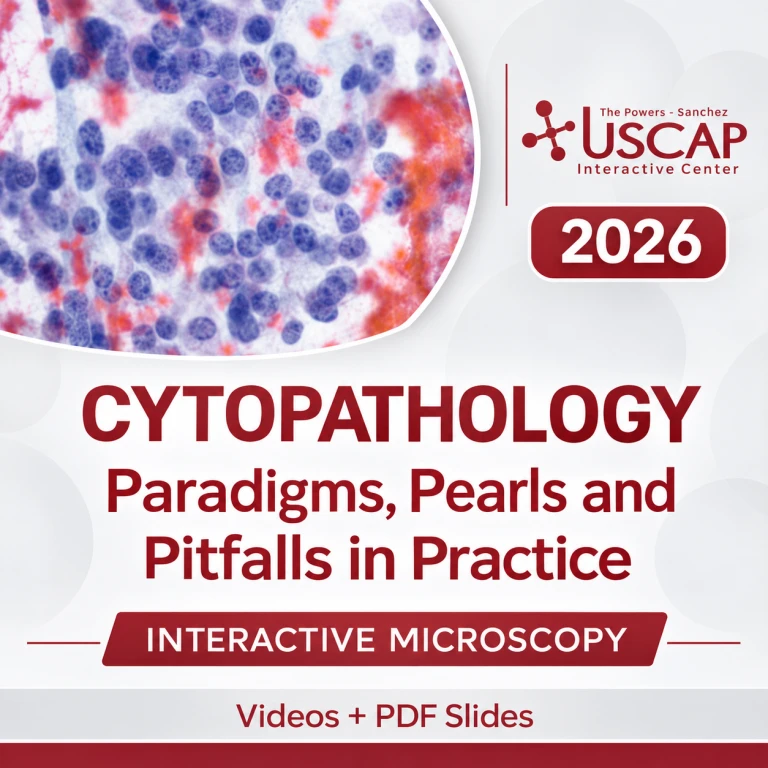
USCAP Cytopathology: Paradigms, Pearls and Pitfalls in Practice 2026
$130.00
USCAP Cytopathology: Paradigms, Pearls and Pitfalls in Practice 2026 features eight expert-led educational sessions focused on diagnostic cytopathology, cytologic classification, differential diagnosis, and practical diagnostic pearls and pitfalls.
The organ-based curriculum covers the urinary tract, kidneys, adrenal glands, serous fluids, pancreas, liver, bile ducts, lungs, mediastinum, soft tissue, thyroid, head and neck, and salivary glands.
Materials: Video recordings and PDF slides · Approximately 27 GB
Original release: June 12, 2026
USCAP Cytopathology: Paradigms, Pearls and Pitfalls in Practice 2026
Cytopathology 2026 | Diagnostic Cytology | Surgical Pathology | Fine Needle Aspiration | Cytologic Classification | USCAP
Target Audience
Cytopathologists, surgical and anatomic pathologists, general pathologists, cytotechnologists, pathology residents and fellows, and other healthcare professionals involved in cytologic diagnosis and specimen interpretation.
Information
USCAP Cytopathology: Paradigms, Pearls and Pitfalls in Practice 2026 is an educational course presented by the United States and Canadian Academy of Pathology (USCAP). Originally released on June 12, 2026, it offers a case-based review of diagnostic cytopathology across multiple organ systems.
The program examines cytomorphology, diagnostic terminology, morphological mimics, differential diagnosis, specimen handling, and ancillary testing. It emphasizes practical interpretation and the correlation of cytologic findings with clinical and laboratory information.
The product description lists video recordings and PDF presentation slides, totaling approximately 27 GB.
Course Details
| Information | Details |
|---|---|
| Course Title | Cytopathology: Paradigms, Pearls and Pitfalls in Practice |
| Organizer | United States and Canadian Academy of Pathology (USCAP) |
| Original Release Date | June 12, 2026 |
| Edition | 2026 |
| Format | On-Demand Educational Recordings |
| Included Materials | Videos + PDF Slides |
| Total File Size | Approximately 27 GB |
| Educational Sessions | 8 |
| Primary Specialty | Cytopathology |
| Additional Focus | Surgical Pathology, Anatomic Pathology, Diagnostic Cytology |
| Language | English |
Learning Objectives
This program reviews how to:
- Apply diagnostic terminology and classify neoplastic and non-neoplastic lesions.
- Recognize cytomorphologic features, diagnostic clues, and common pitfalls across organ systems.
- Develop differential diagnoses and use structured approaches to challenging cases.
- Integrate clinical findings and ancillary test results with cytologic interpretation.
- Review specimen handling and interpretation across urinary, gastrointestinal, pulmonary, endocrine, soft tissue, and head and neck cytopathology.
Complete Course Curriculum – 8 Educational Sessions
1. Urine, Kidney, and Adrenal Cytopathology
Faculty: Adebowale J. Adeniran, MD
Reviews urinary tract, kidney, and adrenal cytopathology, emphasizing cytomorphology, classification, and differential diagnosis.
Educational Focus:
- Urinary tract, kidney, and adrenal cytopathology.
- Cytomorphologic interpretation and classification of neoplastic and non-neoplastic findings.
- Differential diagnosis.
2. Serous Fluid Cytopathology
Faculty: Adebowale J. Adeniran, MD
Examines serous fluid specimens, including cytomorphology, benign and malignant findings, diagnostic mimics, and ancillary testing.
Educational Focus:
- Serous fluid cytology and cytomorphologic features.
- Benign and malignant findings, diagnostic mimics, and cytologic interpretation.
- Considerations for ancillary testing.
3. Pancreas Cytopathology
Faculty: Michelle D. Reid, MD, MSc
Reviews pancreatic cytopathology, lesion classification, diagnostic pitfalls, and the distinction between neoplastic and non-neoplastic findings.
Educational Focus:
- Pancreatic cytopathology and cytologic classification.
- Neoplastic lesions and non-neoplastic abnormalities.
- Diagnostic pitfalls, differential diagnosis, and clinical correlation.
4. Bile Duct and Liver Cytopathology
Faculty: Michelle D. Reid, MD, MSc
Covers bile duct and liver cytology, with emphasis on morphology, lesion classification, and ancillary diagnostic approaches.
Educational Focus:
- Bile duct and liver cytopathology and hepatobiliary lesions.
- Cytomorphology and classification of neoplastic and non-neoplastic conditions.
- Ancillary diagnostic approaches.
5. Lung and Mediastinum Cytopathology
Faculty: Lester J. Layfield, MD
Reviews pulmonary and mediastinal cytology, including tumor classification, diagnostic mimics, and clinical correlation.
Educational Focus:
- Pulmonary and mediastinal cytopathology and cytologic features.
- Diagnostic mimics, tumor classification, and differential diagnosis.
- Clinical and pathological correlation.
6. Soft Tissue Cytopathology
Faculty: Lester J. Layfield, MD
Examines soft tissue lesions, morphological mimics, diagnostic classification, and differential diagnosis.
Educational Focus:
- Soft tissue cytopathology and neoplastic and non-neoplastic lesions.
- Cytologic interpretation, diagnostic classification, and morphological mimics.
- Differential diagnosis.
7. Thyroid Cytopathology
Faculty: William C. Faquin, MD, PhD
Reviews thyroid cytology, reporting terminology, lesion classification, and benign and malignant findings.
Educational Focus:
- Thyroid cytopathology, lesion classification, and diagnostic terminology.
- Cytomorphologic findings in benign and malignant lesions.
- Ancillary testing considerations and differential diagnosis.
8. Head and Neck, and Salivary Gland Cytopathology
Faculty: William C. Faquin, MD, PhD
Covers head and neck and salivary gland cytology, focusing on neoplastic classification, diagnostic pitfalls, and differential diagnosis.
Educational Focus:
- Head and neck and salivary gland cytology.
- Cytomorphologic assessment and neoplastic classification.
- Diagnostic pitfalls, differential diagnosis, and practical interpretation.
Related Medical Resources
Explore related courses in cytopathology and diagnostic cytology:
Educational Notice: This digital product includes recorded educational materials and PDF presentations. It does not include access to the original provider’s learning platform, supervised practical training, CME credits, or certification unless specifically stated in the product package.
Related products
Obstetrics & Gynecology
2023 Surgical Pathology Update: Diagnostic Pearls for the Practicing Pathologist: Vol. VII
Dermatology
Hematology / Oncology

Obstetrics & Gynecology
AIUM Ultrasound of Ankle and Foot Pathology and Therapeutics 2020 (CME VIDEOS)

Neurology / neurosurgery courses