

Oakstone UCSF Neuro and Musculoskeletal Imaging 2026


$35.00
This Product is shared via google drive download link, So please share your correct Gmail id while placing the order .Please note that there are no CME points or certificate associated with this course Samples for Courses Can be found here : Free Samples Here!

Oakstone UCSF Neuro and Musculoskeletal Imaging 2026 (Videos + PDFs)
Master Advanced Neuroradiology and Musculoskeletal Imaging with UCSF Experts
Advance your diagnostic expertise with Oakstone UCSF Neuro and Musculoskeletal Imaging 2026, a comprehensive online continuing medical education (CME) program developed by the renowned faculty of the University of California, San Francisco (UCSF). This advanced course provides an in-depth review of the latest evidence-based practices in neuroradiology and musculoskeletal imaging, helping radiologists confidently interpret complex imaging studies and optimize patient care.
Designed for both subspecialty and general radiologists, the program combines expert-led lectures, challenging case reviews, and practical imaging strategies covering brain, head and neck, spine, peripheral nerves, joints, bones, and soft tissue disorders. Participants will learn how to select optimal imaging protocols, recognize subtle imaging findings, avoid common diagnostic pitfalls, and communicate clinically meaningful reports to referring physicians.
Released in June 2026, this comprehensive educational resource reflects current imaging guidelines, modern MRI and CT protocols, advances in musculoskeletal ultrasound, and contemporary approaches to neurological and orthopedic imaging.
Product Details
- Format: On-Demand Videos + PDFs
- Release Date: June 15, 2026
- Organizer: Oakstone / UCSF
- Language: English
- Educational Level: Intermediate to Advanced
Course Overview
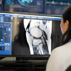
Neuroimaging and musculoskeletal radiology continue to evolve rapidly with advances in MRI techniques, multidetector CT, high-resolution ultrasound, and image-guided diagnostic approaches. Accurate interpretation of these studies plays a critical role in diagnosing neurological emergencies, traumatic injuries, inflammatory diseases, degenerative disorders, congenital abnormalities, and musculoskeletal pathology.
UCSF Neuro and Musculoskeletal Imaging 2026 provides a comprehensive review of clinically relevant topics encountered in everyday radiology practice. Through evidence-based lectures and case-oriented learning, participants will strengthen their understanding of imaging protocols, improve diagnostic confidence, and enhance communication with neurologists, neurosurgeons, orthopedic surgeons, emergency physicians, and other specialists.
The curriculum covers the complete spectrum of neuroradiology and musculoskeletal imaging, including brain disorders, pediatric neuroradiology, head and neck pathology, spinal diseases, sports injuries, arthritis, metabolic bone disorders, musculoskeletal infections, and ultrasound-guided diagnostic applications.
Course Highlights
- Comprehensive neuroradiology review
- Advanced musculoskeletal imaging updates
- Brain, spine, head and neck imaging
- Adult and pediatric neuroradiology
- MRI and CT protocol optimization
- Musculoskeletal ultrasound applications
- Joint imaging and sports injuries
- Spine imaging interpretation
- Bone tumors and metabolic bone disease
- Case-based learning with real-world imaging
- Practical reporting strategies
- Evidence-based diagnostic recommendations
What You’ll Learn
After completing this course, participants will be able to:
- Select appropriate CT and MRI imaging protocols for brain, spine, head and neck, peripheral nerve, and musculoskeletal examinations.
- Recognize imaging features of neurological emergencies requiring urgent diagnosis and intervention.
- Differentiate common and uncommon head and neck masses while recognizing important imaging mimics and post-treatment changes.
- Evaluate headaches using advanced imaging techniques, including assessment of skull base lesions, cranial nerves, sellar, and parasellar abnormalities.
- Interpret imaging findings associated with neonatal brain development, hypoxic injury, autoimmune disorders, and intracranial pressure abnormalities.
- Diagnose spinal degenerative disease, inflammatory disorders, neoplasms, trauma, and postoperative complications using modern imaging approaches.
- Evaluate internal derangements of the shoulder, elbow, hip, knee, ankle, and wrist while avoiding common diagnostic pitfalls.
- Apply musculoskeletal ultrasound as an effective problem-solving tool for challenging orthopedic and sports medicine cases.
- Identify imaging characteristics of infectious, inflammatory, metabolic, and crystal-induced arthropathies.
- Produce clinically relevant radiology reports that improve communication with referring clinicians and optimize patient management.
Comprehensive Topics Covered
Neuroradiology
Review advanced imaging of the brain, cranial nerves, skull base, pituitary region, cerebrovascular disorders, intracranial emergencies, autoimmune diseases, developmental abnormalities, and pediatric neuroradiology using current MRI and CT techniques.
Head & Neck Imaging
Develop practical approaches for evaluating head and neck masses, salivary gland pathology, sinonasal disease, post-treatment imaging, skull base lesions, and common diagnostic mimics.
Spine Imaging
Strengthen your understanding of cervical, thoracic, and lumbar spine imaging, including degenerative disease, trauma, infection, inflammatory conditions, spinal tumors, postoperative imaging, and spinal cord pathology.
Musculoskeletal Imaging
Master interpretation of MRI, CT, and radiographic findings involving the shoulder, elbow, wrist, hand, hip, knee, ankle, and foot. Review common sports injuries, ligament tears, tendon disorders, cartilage lesions, and internal joint derangements.
Musculoskeletal Ultrasound
Learn practical applications of diagnostic musculoskeletal ultrasound for evaluating tendons, ligaments, muscles, nerves, bursae, and superficial soft tissue pathology, while utilizing ultrasound as a complementary imaging modality in difficult diagnostic cases.
Arthritis & Bone Disorders
Review imaging findings associated with inflammatory arthritis, osteoarthritis, crystal arthropathies, metabolic bone disease, osteomyelitis, and musculoskeletal infections using evidence-based diagnostic criteria.
Pediatric Neuroimaging
Explore imaging of neonatal brain development, birth-related injuries, congenital anomalies, autoimmune diseases, and pediatric neurological emergencies.
Imaging Protocol Optimization
Gain practical recommendations for selecting appropriate imaging protocols that maximize diagnostic accuracy while improving workflow efficiency across neuroradiology and musculoskeletal imaging.
Learning Objectives
Upon completion of this educational activity, participants will be able to:
- Recommend appropriate CT and MRI protocols for neuro and musculoskeletal imaging.
- Recognize imaging findings associated with neurological emergencies and complex musculoskeletal disorders.
- Differentiate head and neck masses, skull base lesions, and common imaging mimics.
- Interpret challenging musculoskeletal MRI and ultrasound examinations with greater confidence.
- Apply current imaging strategies for inflammatory, infectious, metabolic, and traumatic disorders.
- Improve diagnostic reporting through structured, clinically meaningful interpretation.
- Utilize musculoskeletal ultrasound as a valuable adjunct for diagnosis and patient management.
- Integrate evidence-based imaging recommendations into daily radiology practice.
Educational Features
- Expert UCSF Faculty
- Comprehensive Neuroradiology Review
- Advanced Musculoskeletal Imaging
- MRI & CT Protocol Optimization
- Case-Based Clinical Learning
- Practical Reporting Strategies
- Evidence-Based Imaging Guidelines
- Challenging Diagnostic Cases
- Musculoskeletal Ultrasound Applications
- On-Demand CME Education
Why This Course Stands Out
UCSF has long been recognized as one of the world’s leading institutions in diagnostic radiology education. This comprehensive program combines the expertise of internationally recognized faculty with practical, case-based instruction that reflects real-world clinical practice.
Unlike narrowly focused imaging courses, Oakstone UCSF Neuro and Musculoskeletal Imaging 2026 integrates neuroradiology and musculoskeletal imaging into a single educational resource, allowing participants to strengthen expertise across two of the most challenging and clinically important areas of diagnostic radiology. The emphasis on optimized imaging protocols, structured reporting, and multidisciplinary communication ensures immediate clinical value for practicing radiologists and trainees alike.
Target Audience
This educational program is ideal for:
- Radiologists
- Neuroradiologists
- Musculoskeletal Radiologists
- General Diagnostic Radiologists
- Radiology Residents
- Radiology Fellows
- Orthopedic Imaging Specialists
- Neurologists
- Neurosurgeons
- Orthopedic Surgeons
- Sports Medicine Physicians
- Emergency Physicians
- Healthcare Professionals interested in advanced diagnostic imaging
Why Choose This Course?
Whether you interpret brain MRI studies, evaluate spinal disorders, diagnose sports injuries, or perform musculoskeletal ultrasound, Oakstone UCSF Neuro and Musculoskeletal Imaging 2026 provides a comprehensive review of current imaging practices supported by evidence-based recommendations and expert clinical experience. Through practical lectures, advanced imaging techniques, and challenging case discussions, participants will strengthen diagnostic accuracy, improve reporting quality, and enhance patient care across a broad range of neurological and musculoskeletal disorders.
+ Topics:
- Imaging of Musculoskeletal Infection – Mini Pathria, MD
- Radiography of Knee Injury – Correlation with CT and MR – Mini Pathria, MD
- MRI of the Knee – Meniscus and Cruciate Ligaments – Kevin Sweetwood, MD
- Arthritis – Beyond Black and White – Kevin Sweetwood, MD
- Metabolic Disorders – Daria Motamedi, MD
- Extensor Mechanism of the Knee – Daria Motamedi, MD
- ACL Graft Reconstruction and its Complications – Daria Motamedi, MD
- Multimodality Evaluation of Tendons of Wrist and Hand – Kevin Sweetwood, MD
- MRI of Shoulder Trauma – Kevin Sweetwood, MD
- Pitfalls in Shoulder MRI – Daria Motamedi, MD
- Hip MRI – Labrum, FAI, and Beyond – Daria Motamedi, MD
- Problem Solved – Applications of MSK US – Daria Motamedi, MD
- MR of the Pelvic Tendons – Mini Pathria, MD
- MR of Muscle Injury – Mini Pathria, MD
- MRI and US Correlation Cases – Kevin Sweetwood, MD
- Ankle and Foot – Commonly Overlooked Injuries – Mini Pathria, MD
- Stress Injury of Bone – Mini Pathria, MD
- Skull Base Lesions – Christine M. Glastonbury, MBBS
- Headache – Do Not Miss Diagnoses – Yi Li, MD
- Top 3 DDX in Head and Neck – Christine M. Glastonbury, MBBS
- Work Up of Non-traumatic Intercranial Hemorrhage – Yi Li, MD
- Imaging the Neonatal Brain – Yi Li, MD
- CSF Pressure Disorders – What the Diagnostic Radiologist Needs to Know – Vinil N. Shah, MD
- Emerging Concepts in Hydrocephalus – Yi Li, MD
- Imaging of Pituitary and Hypothalamic Abnormalities – Yi Li, MD
- Spine Reporting Essentials – How to Add Value – Vinil N. Shah, MD
- Autoimmune Encephalitis – Yi Li, MD
- Spinal Cord Inflammatory Lesions – Vinil N. Shah, MD
- Post Surgical Changes in Head and Neck – Christine M. Glastonbury, MBBS
- Spine Tumor or Mimic – How to Tell Them Apart – Vinil N. Shah, MD
- Salivary Gland Lesions – A Case-Based Review – Christine M. Glastonbury, MBBS
- Spinal Fusion – Postoperative Spine Simplified – Vinil N. Shah, MD
- Pearls and Pitfalls of Head and Neck Tumor Imaging – Christine M. Glastonbury, MBBS
- Challenging Spine Cases – Lessons Learned – Vinil N. Shah, MD
- Errors Are Opportunities – Thinking About QA a Different Way – Christine M. Glastonbury, MBBS
Related products

Cardiology

Critical Care - Emergency medicine
Gulfcoast Ultrasound-Guided Upper Extremity Nerve Blocks for Emergency Medicine

Anesthesiology & pain medicine
GULFCOAST Introduction to Ultrasound-Guided Regional Anesthesia 2019

Critical Care - Emergency medicine
Gulfcoast Pediatric Emergency and Critical Care Ultrasound 2019

Hematology / Oncology
2023 Psychopharmacology in Cancer Care: An Update for Clinicians of All Disciplines


Anesthesiology & pain medicine

Critical Care - Emergency medicine