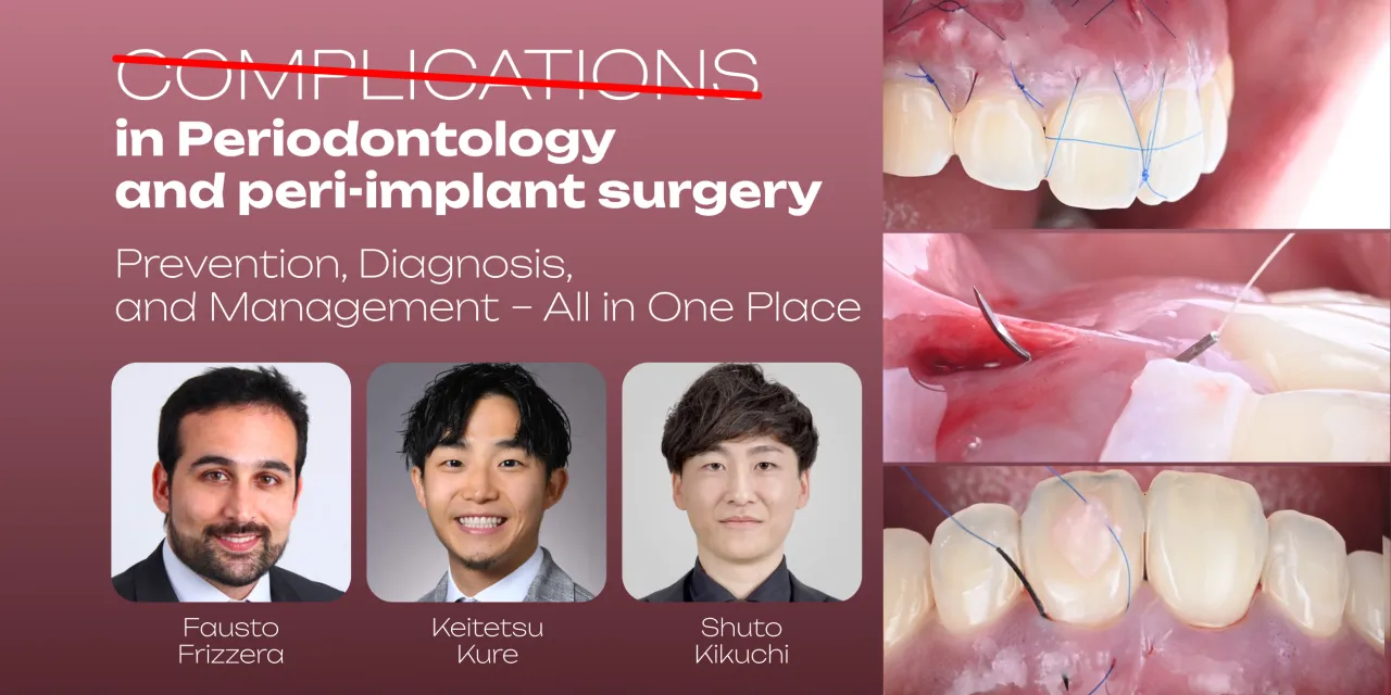
OHI-S Complications in Periodontology and Peri-implant Surgery Prevention, Diagnosis, and Management – All in One Place 5 lessons 4h 33min
$45.00
This Product is shared via google drive download link, So please share your correct Gmail id while placing the order .Please note that there are no CME points or certificate associated with this course Samples for Courses Can be found here : Free Samples Here!
OHI-S Complications in Periodontology and Peri-Implant Surgery
Prevention, Diagnosis, and Management – All in One Place
4 Lessons + 1 Free | 4 Hours 33 Minutes | 4 CE Credits
Develop a structured approach to preventing, diagnosing, and managing periodontal and peri-implant surgical complications with OHI-S Complications in Periodontology and Peri-implant Surgery: Prevention, Diagnosis, and Management – All in One Place.
Presented by Fausto Frizzera, Keitetsu Kure, and Shuto Kikuchi, this advanced clinical program brings together three major areas that are often taught separately:
- Peri-implant soft-tissue complications
- Peri-implantitis and peri-implant bone loss
- Regenerative and mucogingival surgical complications
The course reviews how complications can be evaluated according to:
- Etiology
- Tissue phenotype
- Implant position
- Soft-tissue volume
- Bone-defect morphology
- Surgical technique
- Prosthetic factors
- Wound healing
- Patient risk
- Maintenance
Major topics include:
- Peri-implant recession
- Soft-tissue volume loss
- Papilla deficiencies
- Peri-implant plastic surgery
- Implant removal and site reconstruction
- Peri-implantitis
- Implant-surface decontamination
- Regenerative peri-implant surgery
- Resective peri-implant surgery
- Guided Bone Regeneration – GBR
- Membrane exposure
- Graft infection
- Partial and complete graft loss
- Secondary bone reconstruction
- Gingival recession
- Miller and Cairo classifications
- Root coverage
- Papilla reconstruction
- Tunneling
- Connective tissue grafting
- Microsurgical suturing
- Supportive peri-implant therapy
The central clinical framework is:
Prevent → Identify the Complication → Determine the Cause → Classify the Defect → Select the Appropriate Intervention → Reevaluate → Maintain
Course Details
- Course: Complications in Periodontology and Peri-implant Surgery Prevention, Diagnosis, and Management – All in One Place
- Provider: OHI-S
- Faculty: Fausto Frizzera, Keitetsu Kure, Shuto Kikuchi
- Official Structure: 4 Lessons + 1 Free
- Total Presentations: 5
- Official Duration: 4 Hours 33 Minutes
- Language: English
- Official OHI-S CE: 4 CE Credits
- Primary Specialty: Periodontology
- Major Secondary Specialty: Implantology
- Additional Specialty: Oral & Maxillofacial Surgery
- Clinical Level: Advanced
- Core Focus: Periodontal, Peri-Implant, Regenerative & Soft-Tissue Complications
Course Overview
Complications following periodontal or implant surgery do not all arise from the same mechanism.
A failing result may originate from:
Biological Factors
- Infection
- Tissue inflammation
- Poor wound healing
- Peri-implant disease
Surgical Factors
- Flap design
- Closure tension
- Graft instability
- Membrane handling
- Surgical trauma
Prosthetic Factors
- Implant position
- Emergence profile
- Restorative contours
- Cleansability
Patient Factors
- Smoking
- Systemic disease
- Oral hygiene
- Compliance
A useful complication-management process therefore begins with:
Why Did This Happen?
before asking:
How Do I Correct It?
PERI-IMPLANT SOFT-TISSUE COMPLICATIONS
Soft-tissue problems around implants can affect:
- Aesthetics
- Hygiene
- Biological stability
- Patient satisfaction
- Restorative integration
Common presentations addressed in the course include:
- Soft-tissue volume loss
- Peri-implant recession
- Papilla deficiency
- Aesthetic compromise
These problems should be assessed according to both surgical and prosthetic factors.
LESSON 1
Peri-Implant Soft Tissue Defects: Diagnosis and Complications
Instructor: Fausto Frizzera
Duration: 11 Minutes
Format: Free Clinical Video
The opening lesson provides a focused framework for identifying peri-implant soft-tissue problems.
Topics include:
- Clinical diagnosis of peri-implant soft-tissue defects
- Assessment of soft-tissue volume loss
- Peri-implant soft-tissue recession
- Loss of interproximal papilla
- Aesthetic compromise
- Surgical technique-related complications
- Prosthetic-driven tissue alterations
- Prevention of aesthetic failures around implants
Soft-Tissue Volume Loss
A peri-implant site may lose soft-tissue volume for several reasons.
Potential contributors include:
- Pre-existing phenotype
- Implant position
- Tissue remodeling
- Surgical procedures
- Restorative design
- Inadequate tissue support
Diagnosis should therefore include the complete implant-restorative environment.
Peri-Implant Recession
Peri-implant recession can expose:
- Implant components
- Abutment surfaces
- Restorative margins
The problem may be especially significant in the aesthetic zone.
Management should begin by identifying:
- Implant position
- Buccal tissue thickness
- Bone support
- Restorative emergence
- Interproximal tissue condition
Papilla Deficiency
Loss of the interproximal papilla may produce:
- Black triangles
- Aesthetic compromise
- Food retention
- Patient dissatisfaction
Papilla reconstruction can be technically challenging because soft-tissue appearance depends on the underlying anatomy and restorative relationships.
Surgically Driven vs. Prosthetically Driven Complications
An important concept is distinguishing:
Surgical Contributors
such as tissue loss or implant-position problems
from:
Prosthetic Contributors
such as inappropriate emergence profile or restorative contour.
Correcting soft tissue without addressing the restorative cause may not provide a stable result.
LESSON 2
Management of Soft Tissue Complications Around Dental Implants: Prevention, Diagnosis, and Treatment
Instructor: Fausto Frizzera
Duration: 54 Minutes
This lesson expands from diagnosis into reconstructive decision-making.
Topics include:
- Classification of peri-implant soft-tissue alterations
- Prevention of soft-tissue complications
- Aesthetic implant sites
- Treatment indications
- Timing
- Prognosis
- Peri-implant plastic surgery
- Soft-tissue reconstruction
- Implant failure
- Implant removal
- Site preservation
- Reconstruction following implant removal
Prevention Before Reconstruction
The strongest treatment for a complication is often preventing the complication from developing.
Risk assessment may include:
- Tissue phenotype
- Bone anatomy
- Implant position
- Implant depth
- Restorative plan
- Emergence profile
- Patient hygiene
A useful approach is:
Plan Bone + Implant + Soft Tissue + Prosthesis Together
Peri-Implant Plastic Surgery
Soft-tissue reconstructive procedures may be considered when the implant site demonstrates:
- Recession
- Volume deficiency
- Aesthetic compromise
- Insufficient soft-tissue architecture
Treatment selection depends on the specific defect.
Treatment Timing
The clinician must decide:
- Treat immediately?
- Allow tissue maturation?
- Correct the prosthesis first?
- Remove the implant?
- Reconstruct the site before reimplantation?
There is no single timing protocol for every complication.
Prognosis
Before attempting reconstruction, clinicians should determine whether the implant itself is compatible with a successful final result.
Factors include:
- Implant position
- Bone support
- Biological condition
- Prosthetic feasibility
A poorly positioned or severely compromised implant may not always be best managed by adding more soft tissue.
Implant Removal
The official curriculum specifically includes:
Implant removal, site preservation, and soft-tissue reconstruction following implant failure.
This is an important clinical principle:
Saving the Implant Is Not Always the Same as Saving the Treatment.
In selected cases, removal and reconstruction may offer a more appropriate pathway.
PERI-IMPLANTITIS
Peri-implantitis is a plaque-associated pathological condition involving inflammation around a dental implant with progressive loss of supporting bone.
Management requires assessment of:
- Clinical inflammation
- Probing findings
- Radiographic bone loss
- Implant position
- Defect anatomy
- Prosthetic design
- Patient risk factors
LESSON 3
Surgical Management of Peri-Implantitis: Contemporary Approaches to Implant Complications
Instructor: Fausto Frizzera
Duration: 1 Hour 6 Minutes
This lesson focuses on advanced surgical management of peri-implant disease.
Topics include:
- Clinical diagnosis of peri-implantitis
- Radiographic diagnosis
- Implant-surface decontamination
- Peri-implant bone-defect classification
- Defect morphology
- Regenerative surgery
- Resective surgery
- Treatment indications
- Long-term maintenance
- Supportive peri-implant therapy
Clinical & Radiographic Diagnosis
Peri-implantitis should be assessed using both:
Clinical Findings
and:
Radiographic Findings
because neither alone provides the entire disease picture.
The clinician should evaluate the progression and morphology of bone loss rather than simply observing that bone loss exists.
Defect-Driven Decision-Making
One of the strongest concepts in the course is:
Treat the Defect According to Its Morphology.
The anatomy of the peri-implant bone defect may influence whether the clinician considers:
- Regenerative treatment
- Resective treatment
- Combined treatment
- Implant removal
Implant-Surface Decontamination
Implant-surface management is a critical component of peri-implantitis surgery.
The course discusses contemporary decontamination approaches as part of the overall treatment pathway.
MedicalAmboss should avoid implying that any one decontamination method universally eliminates peri-implantitis.
The appropriate approach depends on:
- Implant surface
- Access
- Defect morphology
- Disease severity
- Surgical strategy
Regenerative Peri-Implant Surgery
Regenerative approaches may be considered in selected defects with favorable anatomy.
Potential objectives include:
- Defect reconstruction
- Bone fill
- Improved tissue support
However, regeneration should not be presented as guaranteed.
Resective Surgery
Resective approaches may be indicated where defect anatomy does not support a predictable regenerative approach.
Clinical decisions should consider:
- Bone morphology
- Implant position
- Surface accessibility
- Aesthetic demands
- Hygiene access
Regenerative vs. Resective Treatment
A useful conceptual pathway is:
Diagnose
↓
Assess Defect Morphology
↓
Evaluate Implant Position and Prognosis
↓
Determine Regenerative / Resective / Combined / Removal Strategy
↓
Decontaminate
↓
Reconstruct or Reshape as Indicated
↓
Maintenance
Supportive Peri-Implant Therapy
Surgery alone is not sufficient for long-term control.
Maintenance may include:
- Plaque control
- Professional monitoring
- Peri-implant probing
- Bleeding assessment
- Radiographic monitoring when indicated
- Prosthetic hygiene evaluation
The objective is to reduce the risk of recurrent disease.
GUIDED BONE REGENERATION – GBR
GBR is an important reconstructive technique in implant dentistry.
However, complications can occur at multiple stages:
- Before surgery
- During surgery
- During wound healing
- During graft maturation
The course focuses specifically on understanding why GBR fails.
LESSON 4
Rethinking GBR Success: Managing Failures and Achieving Predictable Bone Regeneration
Instructor: Keitetsu Kure
Duration: 1 Hour 2 Minutes
This lesson provides a systematic framework for GBR failure.
Topics include:
- Biological principles of bone regeneration
- GBR indications
- GBR objectives
- Wound healing
- Space maintenance
- Regenerative risk factors
- Surgical causes of failure
- Patient-related causes
- Material-related causes
- Biological causes
- Mechanical causes
- Early vs. late complications
- Partial vs. complete graft loss
- Membrane exposure
- Soft-tissue dehiscence
- Infection
- Radiographic assessment
- Intraoperative assessment
- Failed GBR management
- Immediate vs. delayed intervention
- Secondary grafting
- Site reconstruction
- Retreatment criteria
- Prevention
Why GBR Fails
GBR failure is often multifactorial.
The official curriculum organizes causes into several major categories.
Surgical Factors
- Flap design
- Tension-free closure
- Membrane handling
- Infection control
Patient-Related Factors
- Systemic conditions
- Smoking
- Oral hygiene
- Compliance
Material-Related Factors
- Graft selection
- Membrane characteristics
- Stability
- Fixation
Biological / Mechanical Factors
- Healing environment
- Mechanical instability
- Loss of regenerative space
This framework is valuable because it moves complication management away from blaming one isolated material or technique.
Wound Closure
Primary wound stability is central to GBR.
The curriculum specifically highlights:
Tension-Free Closure
because excessive flap tension can contribute to:
- Dehiscence
- Membrane exposure
- Graft contamination
Space Maintenance
Successful GBR requires maintenance of an appropriate regenerative space.
Collapse or instability can compromise the intended reconstruction.
This makes:
- Membrane design
- Graft stability
- Fixation
important components of the overall mechanical environment.
Early vs. Late GBR Complications
The timing of a complication can influence management.
Early Complications
may occur during initial healing.
Late Complications
may occur after a period of graft maturation.
The clinician should consider:
- Timing
- Exposure
- Infection
- Graft stability
- Clinical findings
before deciding how aggressively to intervene.
Partial vs. Complete Graft Loss
GBR failure is not necessarily binary.
A case may demonstrate:
- Partial compromise
- Partial graft loss
- Complete failure
Management should be based on the actual clinical and radiographic condition rather than automatically removing every graft.
Membrane Exposure
Membrane exposure is one of the major complications covered.
Management depends on factors including:
- Membrane type
- Timing
- Infection
- Soft-tissue condition
- Graft stability
The course reviews how to evaluate these compromised regenerative sites.
Infection
Infection can severely affect regenerative outcomes.
The appropriate response should consider:
- Extent of infection
- Implant involvement
- Membrane status
- Graft condition
- Patient systemic status
Antimicrobial management should follow appropriate professional guidance and patient-specific indications.
Failed GBR: Immediate vs. Delayed Intervention
The clinician may need to choose between:
Immediate Intervention
during the complication episode
and:
Delayed Reconstruction
after healing and site reassessment.
The official curriculum specifically compares these strategies.
Secondary Grafting
A failed regenerative procedure does not always mean that further reconstruction is impossible.
The course includes:
- Secondary grafting
- Site reconstruction
- Criteria for retreatment
A second procedure should address the cause of the first failure rather than simply repeating the original protocol unchanged.
ROOT COVERAGE & PAPILLA RECONSTRUCTION
The final lesson moves from implant complications into advanced periodontal plastic surgery.
This includes:
- Gingival recession
- Root coverage
- Papilla deficiency
- Tunneling
- CTG
- Microsurgical suturing
LESSON 5
Root Coverage and Papilla Reconstruction: Advanced Surgical Techniques and Clinical Protocols
Instructor: Shuto Kikuchi
Duration: 1 Hour 18 Minutes
This is the longest individual lesson in the course and focuses on advanced mucogingival reconstruction.
Topics include:
- Miller recession classification
- Cairo recession classification
- Root-coverage diagnosis
- Treatment planning
- Incision design
- Flap elevation
- Microsurgical suturing
- Suprabony defects
- Intrabony defects
- Partial-thickness flaps
- Split-full-split flaps
- Supracrestal periodontal reconstruction
- Interproximal tunneling
- Customized connective tissue grafts
- Papilla reconstruction
- CTG
- Interproximal sling sutures
- Double-crossed sutures
- Complication prevention
- Clinical cases
Gingival Recession Classification
The course reviews both:
Miller Classification
and:
Cairo Classification
These frameworks help clinicians describe recession defects and assess treatment possibilities.
Root-Coverage Treatment Planning
The decision to perform root coverage should consider:
- Recession anatomy
- Interproximal attachment
- Tissue phenotype
- Keratinized tissue
- Tooth position
- Patient expectations
The choice of technique should follow diagnosis.
Partial-Thickness Flaps
Partial-thickness approaches preserve selected tissue layers while mobilizing the flap.
They require precise anatomical knowledge and surgical control.
Split-Full-Split Flap
The official course includes the:
Split–Full–Split / Mixed-Thickness Flap
concept.
Different flap thicknesses may be used at different portions of the surgical site according to:
- Anatomy
- vascular supply
- flap mobility
- grafting strategy
Tunneling Technique
Interproximal tunneling aims to mobilize soft tissue while minimizing external incisions in selected cases.
The curriculum includes:
- Interproximal tunneling
- Customized connective tissue grafts
- Papilla reconstruction
Connective Tissue Graft – CTG
CTG is a major reconstructive technique in periodontal plastic surgery.
The program discusses CTG in relation to:
- Root coverage
- Papilla reconstruction
- Tissue augmentation
Clinical success depends on:
- Graft design
- Recipient-site preparation
- Vascular supply
- Flap stability
- Suturing
Papilla Reconstruction
Papilla reconstruction remains one of the most challenging soft-tissue procedures.
The clinician must assess:
- Interproximal bone
- Contact point
- Tissue availability
- Scar formation
- Tooth / implant relationships
Treatment should therefore be approached with realistic expectations.
Microsurgical Suturing
The official curriculum includes:
- Interrupted interproximal sling sutures
- Double-crossed sutures
The objective of microsurgical suturing is to support:
- Precise tissue positioning
- Primary stability
- Wound adaptation
rather than simply closing the surgical site.
Supracrestal Tissue Reconstruction
The lesson includes supracrestal or supra-alveolar periodontal tissue reconstruction protocols.
These techniques are advanced and should be understood within:
- Defect classification
- biological limits
- periodontal anatomy
PERI-IMPLANT SOFT-TISSUE DEFECT vs. PERI-IMPLANTITIS
These should not be conflated.
Soft-Tissue Defect
May primarily involve:
- recession
- volume deficiency
- papilla loss
without necessarily representing active peri-implantitis.
Peri-Implantitis
Involves an inflammatory disease process with progressive supporting bone loss.
The treatment pathway differs substantially.
BIOLOGICAL vs. AESTHETIC COMPLICATION
Another key distinction is:
Biological Complication
Examples:
- peri-implantitis
- infection
- progressive bone loss
Aesthetic / Morphologic Complication
Examples:
- recession
- soft-tissue deficiency
- papilla loss
A patient may have either or both.
SURGICAL vs. PROSTHETIC CAUSE
Peri-implant tissue problems can result from:
Surgery
- implant position
- bone deficiency
- tissue damage
Prosthetics
- emergence profile
- contour
- cleansability
The definitive treatment should address the dominant cause.
Comprehensive Complication Algorithm
A useful framework is:
Step 1 — Identify the Problem
- Soft tissue?
- Bone?
- Infection?
- Graft failure?
- Aesthetic defect?
↓
Step 2 — Determine Etiology
- Surgical?
- Biological?
- Prosthetic?
- Patient-related?
- Material-related?
↓
Step 3 — Assess Prognosis
- Can the implant be maintained?
- Can tissue be reconstructed?
- Is removal required?
↓
Step 4 — Select Treatment
- Soft-tissue reconstruction
- Peri-implant surgery
- GBR revision
- Implant removal
- Root coverage
- Papilla reconstruction
↓
Step 5 — Maintenance
- Hygiene
- Follow-up
- Supportive periodontal/peri-implant therapy
Major Topics Covered
- Periodontology Complications
- Peri-Implant Surgery Complications
- Implant Complications
- Peri-Implant Soft Tissue
- Peri-Implant Recession
- Papilla Loss
- Peri-Implantitis
- Implant Surface Decontamination
- Peri-Implant Bone Loss
- Regenerative Surgery
- Resective Surgery
- Guided Bone Regeneration
- GBR Failure
- Membrane Exposure
- Graft Infection
- Bone Graft Failure
- Secondary Bone Grafting
- Soft Tissue Dehiscence
- Periodontal Plastic Surgery
- Gingival Recession
- Miller Classification
- Cairo Classification
- Root Coverage
- Papilla Reconstruction
- Tunneling
- Connective Tissue Graft
- CTG
- Microsurgical Suturing
- Implant Removal
- Supportive Peri-Implant Therapy
Who Should Take This Course?
The strongest target audience includes:
- Periodontists
- Implantologists
- Oral and Maxillofacial Surgeons
- Periodontology Residents
- Implant Dentistry Residents
- Advanced General Dentists with formal surgical training
The course can also be highly relevant to:
- Prosthodontists
- Restorative Dentists involved in implant rehabilitation
because prosthetic design can contribute to or complicate peri-implant soft-tissue problems.
Primary Audience
The strongest primary audience is:
Periodontists + Implantologists
because the program combines:
- Mucogingival surgery
- Peri-implantitis
- Soft-tissue augmentation
- GBR
- Regenerative surgery
Oral and maxillofacial surgeons represent an important advanced surgical audience.
Restorative dentists should be considered a secondary interdisciplinary audience, particularly for understanding prosthetic contributors to implant complications.
Why This Course Is Useful
The course covers five different levels of complications.
1. Soft-Tissue Diagnosis
- Recession
- Volume loss
- Papilla deficiency
2. Soft-Tissue Reconstruction
- Peri-implant plastic surgery
- Implant-removal/site reconstruction
3. Peri-Implant Disease
- Diagnosis
- Decontamination
- Regenerative vs. resective surgery
4. Bone Regeneration Failure
- GBR exposure
- Infection
- Graft loss
- Secondary reconstruction
5. Mucogingival Reconstruction
- Root coverage
- Tunneling
- CTG
- Papilla reconstruction
The complete educational pathway is:
Soft Tissue → Disease → Bone → Reconstruction → Maintenance
Important Outcome Positioning
The original OHI-S language refers to complications as potentially “predictable, treatable conditions.”
For MedicalAmboss, a safer formulation is:
“The course provides structured protocols for analyzing complications and selecting appropriate management strategies.”
Avoid claims such as:
- All complications are predictable.
- Every failed GBR can be regenerated.
- Peri-implantitis can always be resolved surgically.
- Papilla reconstruction is predictable in every patient.
- CTG guarantees complete root coverage.
Clinical results depend on:
- Defect morphology
- Patient health
- implant position
- tissue phenotype
- smoking
- infection
- surgical technique
- prosthetic design
- healing
- maintenance
Important Surgical Training Notice
This is an advanced surgical course.
Procedures discussed include:
- Peri-implant plastic surgery
- Peri-implantitis surgery
- GBR
- secondary bone grafting
- implant removal
- CTG
- tunneling
- papilla reconstruction
- microsurgical suturing
Recorded education can support:
- diagnosis
- treatment planning
- complication recognition
- surgical sequencing
but does not replace:
- formal periodontal/implant surgical training
- hands-on instruction
- supervised clinical experience
- patient-specific diagnosis
- emergency and complication-management competency
- appropriate professional credentialing
Long-Term Maintenance
Complication management does not end after surgery.
Long-term stability requires:
- Biofilm control
- Periodontal maintenance
- Peri-implant maintenance
- Reassessment
- Prosthetic hygiene access
- Risk-factor management
- Early identification of recurrent disease
A successful reconstructive result that cannot be adequately maintained may remain at risk.
Original OHI-S CE Information
The current official OHI-S course is listed as:
4 Lessons + 1 Free
with:
4 Hours 33 Minutes
and:
4 CE Credits
Official CE eligibility applies to eligible participation through the original OHI-S educational activity and completion of its requirements.
An independently distributed MedicalAmboss recording package should not automatically be advertised as including:
- OHI-S CE Credits
- OHI-S Certificate
unless official participation and CE eligibility are specifically included.
Product Summary
- Product: Complications in Periodontology and Peri-implant Surgery Prevention, Diagnosis, and Management – All in One Place
- Provider: OHI-S
- Faculty: Fausto Frizzera, Keitetsu Kure, Shuto Kikuchi
- Official Structure: 4 Lessons + 1 Free
- Total Presentations: 5
- Official Duration: 4h 33min
- Language: English
- Official OHI-S CE: 4 Credits
- Primary Specialty: Periodontology
- Major Secondary Specialty: Implantology
- Additional Specialty: Oral & Maxillofacial Surgery
- Core Focus: Soft-Tissue Complications, Peri-Implantitis, GBR Failure & Mucogingival Reconstruction
4. Short Description
OHI-S Complications in Periodontology and Peri-implant Surgery: Prevention, Diagnosis, and Management – All in One Place is an advanced 4h 33min surgical course by Fausto Frizzera, Keitetsu Kure, and Shuto Kikuchi, officially structured as 4 lessons + 1 free clinical video.
The program covers peri-implant soft-tissue recession and volume loss, papilla deficiencies, peri-implant plastic surgery, implant removal and site reconstruction, peri-implantitis diagnosis, implant-surface decontamination, regenerative and resective surgery, GBR failure, membrane exposure, infection, secondary grafting, Miller and Cairo recession classifications, root coverage, tunneling, connective tissue grafting, papilla reconstruction, microsurgical suturing, and long-term supportive peri-implant therapy.
Topics
Lesson 1. Peri-Implant Soft Tissue Defects: Diagnosis and Complications
-
Clinical diagnosis of peri-implant soft tissue defects
-
Assessment of soft tissue volume loss
-
Peri-implant soft tissue recession
-
Loss of interproximal papilla and esthetic compromise
-
Surgical technique-related complications
-
Prosthetic-driven soft tissue alterations
-
Prevention of esthetic failures around implants
Lesson 2. Management of Soft Tissue Complications Around Dental Implants: Prevention, Diagnosis, and Treatment
-
Diagnosis and classification of peri-implant soft tissue alterations
-
Prevention of soft tissue complications in esthetic implant sites
-
Clinical decision-making: treatment indications, timing, and prognosis
-
Peri-implant plastic surgery techniques for soft tissue reconstruction
-
Implant removal, site preservation, and soft tissue reconstruction following implant failure
Lesson 3. Surgical Management of Peri-Implantitis: Contemporary Approaches to Implant Complications
-
Clinical and radiographic diagnosis of peri-implantitis
-
Implant surface decontamination techniques
-
Classification and management of peri-implant bone defects
-
Regenerative surgical approaches for peri-implantitis
-
Resective surgical procedures and clinical indications
-
Long-term maintenance protocols and supportive peri-implant therapy
Lesson 4. Root Coverage and Papilla Reconstruction: Advanced Surgical Techniques and Clinical Protocols
-
Classification of gingival recessions: Miller and Cairo system
-
Clinical diagnosis and treatment planning for root coverage procedures
-
Incision design, flap elevation, and microsurgical suturing principles
-
Management of suprabony and intrabony periodontal defects
-
Partial-thickness and split-full-split (mixed-thickness) flap techniques
-
Supracrestal (supra-alveolar) periodontal tissue reconstruction protocols
-
Interproximal tunneling with customized connective tissue grafts
-
Connective tissue graft (CTG) techniques for papilla reconstruction
-
Microsurgical suturing techniques: interrupted interproximal sling and double-crossed sutures
-
Prevention and management of complications in root coverage surgery
-
Step-by-step analysis of clinical cases and treatment outcomes
Lesson 5. Rethinking GBR Success: Managing Failures and Achieving Predictable Bone Regeneration
-
Biological principles of bone regeneration
-
Indications and objectives of GBR
-
Factors influencing predictable regenerative outcomes
-
Principles of wound healing and space maintenance
-
Aetiology of GBR failures:
-
Surgical factors: flap design, tension-free closure, membrane handling, and infection control
-
Patient-related risk factors: systemic conditions, smoking, oral hygiene, and compliance
-
Material-related factors: graft selection, membrane characteristics, stability, and fixation
-
Biological and mechanical causes of regenerative failure
-
-
Classification and diagnosis of GBR complications:
-
Early versus late complications
-
Partial versus complete graft loss
-
Soft tissue dehiscence and membrane exposure
-
Clinical and radiographic assessment of compromised regenerative sites
-
-
Review of representative GBR failure cases
-
Identification of surgical errors and contributing risk factors
-
Clinical decision-making based on radiographic and intraoperative findings
-
Management of failed GBR procedures
-
Immediate versus delayed intervention strategies
-
Management of membrane exposure and infection
-
Secondary grafting techniques and site reconstruction
-
Criteria for retreatment and achieving predictable outcomes
-
Prevention and long-term success
Related products

DENTAL
Sinus Grafting for Implant Reconstruction (Pikos Online MasterClass Series) – Michael A. Pikos
DENTAL