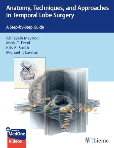
Anatomy, Techniques, and Approaches in Temporal Lobe Surgery: A Step-by-Step Guide (Original PDF from Publisher+Videos)

Original price was: $226.30.$25.00Current price is: $25.00.
This Product is shared via google drive download link, So please share your correct Gmail id while placing the order .Please note that there are no CME points or certificate associated with this course Samples for Courses Can be found here : Free Samples Here!
Anatomy, Techniques, and Approaches in Temporal Lobe Surgery: A Step-by-Step Guide (Original PDF from Publisher + Videos)
Temporal Lobe Surgery | Temporal Lobe Epilepsy Surgery | Neurosurgical Anatomy | Epilepsy Surgery | Surgical Approaches | Cadaveric Dissection | 3D Neurosurgical Anatomy | Operative Videos
Information
Anatomy, Techniques, and Approaches in Temporal Lobe Surgery: A Step-by-Step Guide (Original PDF from Publisher + Videos) is a comprehensive neurosurgical resource dedicated to the anatomy, operative approaches, and step-by-step surgical techniques used in the management of temporal lobe epilepsy (TLE).
Edited by Ali Tayebi Meybodi, Mark C. Preul, Kris A. Smith, and Michael T. Lawton, the book combines detailed anatomical instruction with cadaveric dissection, surgically oriented illustrations, high-resolution three-dimensional images, and narrated procedural videos.
The temporal lobe is one of the most anatomically and functionally complex regions of the brain. Safe surgery requires detailed understanding of cortical and subcortical anatomy, vascular relationships, meningeal structures, white matter pathways, and functionally important neighboring regions.
This resource is designed to bridge the gap between didactic neuroanatomy and practical operative training by presenting modern temporal lobe epilepsy procedures in a systematic, step-by-step format.
Product Details
- Title: Anatomy, Techniques, and Approaches in Temporal Lobe Surgery
- Subtitle: A Step-by-Step Guide
- Edition: 1st Edition
- Edition Year: 2027
- Release Date: October 14, 2026
- Copyright Year: 2026
- Format Available Here: Original Publisher PDF + Videos
- Publisher: Thieme
- Editors: Ali Tayebi Meybodi, Mark C. Preul, Kris A. Smith, Michael T. Lawton
- PDF Length: 548 pages
- Images: 997
- Language: English
- Specialty: Neurosurgery / Epilepsy Surgery / Neuroanatomy
- ISBN (PDF): 9781684206353
Thieme lists the PDF edition as 548 pages with 997 images, released on October 14, 2026, and identifies it as the 1st Edition 2027.
Target Audience
This resource is particularly relevant for:
- Neurosurgeons
- Epilepsy Surgeons
- Functional Neurosurgeons
- Cerebrovascular Neurosurgeons
- Skull Base Neurosurgeons
- Neurosurgical Residents
- Neurosurgical Fellows
- Epileptologists
- Neurologists involved in epilepsy surgery programs
- Neuroanatomy Fellows
- Surgical Neuroanatomy Researchers
- Clinicians training in temporal lobe surgery
Program Overview
The book provides a structured progression from foundational principles to detailed operative technique.
The content begins with:
- History of temporal lobe epilepsy surgery
- Functional anatomy of the temporal lobe
- Surgical anatomy of the temporal lobe
It then progresses into seven dedicated procedural chapters that systematically demonstrate contemporary surgical approaches used in temporal lobe epilepsy surgery.
Each procedural chapter integrates:
- Surgical anatomy
- Operative orientation
- Step-by-step technique
- Surgically oriented views
- Cadaveric dissections
- Detailed figures
- High-resolution 3D images
- Narrated videos
Thieme specifically describes the book as combining three foundational chapters with seven procedural chapters covering current TLE surgical approaches.
Key Learning Areas
This resource supports advanced study of:
- Temporal lobe anatomy
- Temporal lobe epilepsy surgery
- Surgical corridors
- Cortical anatomy
- Meningeal anatomy
- Vascular anatomy
- Neural anatomy
- White matter anatomy
- Operative landmarks
- Microsurgical dissection
- Epilepsy surgery planning
- Safe temporal lobe resection
- Cadaveric surgical training
- Surgical orientation
- Three-dimensional neuroanatomy
Major Topics Covered
Temporal Lobe Epilepsy Surgery
Temporal lobe epilepsy is among the most common surgically treated forms of focal epilepsy.
Surgical management requires precise understanding of:
- Epileptogenic anatomy
- Temporal lobe structures
- Mesial temporal anatomy
- Neurovascular relationships
- Functional boundaries
- Surgical corridors
- Potential neurological complications
The book focuses specifically on resection techniques used in the operative management of TLE.
History of Temporal Lobe Epilepsy Surgery
The introductory chapter reviews the evolution of temporal lobe surgery and provides historical context for contemporary techniques.
Understanding the development of epilepsy surgery helps learners appreciate:
- How operative approaches evolved
- Why certain techniques became preferred
- How anatomical knowledge shaped modern surgery
- How current procedures build on earlier surgical experience
Functional Anatomy of the Temporal Lobe
Safe temporal lobe surgery requires more than recognition of gross anatomical structures.
Functional anatomy is critical because the temporal lobe contributes to:
- Language
- Memory
- Auditory processing
- Visual processing
- Emotional function
- Higher cognitive processes
The functional anatomy chapter helps learners relate surgical corridors to clinically important brain regions.
Surgical Anatomy of the Temporal Lobe
The dedicated surgical anatomy section provides the anatomical foundation for the procedural chapters.
Key anatomical domains include:
- Cortical anatomy
- Sulci and gyri
- Mesial temporal structures
- Ventricular relationships
- Vascular anatomy
- Meningeal structures
- Neural structures
- White matter pathways
- Skull base relationships
Cadaveric Dissection
A major strength of the book is its extensive use of detailed cadaveric dissections.
The dissections focus on:
- Bony anatomy
- Meningeal anatomy
- Vascular structures
- Neural structures
- Surgical corridors
- Deep anatomical relationships
Thieme specifically highlights these dissections as a core feature for understanding temporal lobe surgical anatomy.
Bony Anatomy
Temporal lobe surgery requires familiarity with the surrounding cranial architecture.
Important relationships may include:
- Temporal bone
- Middle cranial fossa
- Skull base
- Sphenoid region
- Bony surgical landmarks
Understanding these structures helps orient the surgeon during craniotomy and deeper dissection.
Meningeal Anatomy
The meninges form important anatomical boundaries and corridors during neurosurgery.
Surgical planning may require understanding of:
- Dura
- Arachnoid
- Subarachnoid spaces
- Cisternal anatomy
- Meningeal relationships to vessels and nerves
Vascular Anatomy
Temporal lobe surgery carries significant vascular considerations.
Relevant structures may include:
- Middle cerebral artery branches
- Posterior cerebral circulation
- Temporal arterial branches
- Venous drainage
- Deep vessels
- Perforating arteries
Precise vascular knowledge is essential for minimizing ischemic and hemorrhagic complications.
Neural Anatomy
The surgical anatomy of the temporal lobe includes important neural structures and pathways.
The procedural material helps learners understand the spatial relationship between:
- Temporal neocortex
- Mesial temporal structures
- Limbic structures
- Adjacent cortical regions
- Deep neural structures
- White matter pathways
Mesial Temporal Anatomy
Mesial temporal structures are particularly important in epilepsy surgery.
Relevant anatomy may include:
- Hippocampal region
- Amygdalar region
- Parahippocampal structures
- Temporal horn
- Adjacent cisternal anatomy
A detailed three-dimensional understanding of these regions is fundamental for safe surgical resection.
Surgical Approaches to Temporal Lobe Epilepsy
The seven procedural chapters provide systematic, step-by-step demonstrations of current surgical techniques used in TLE surgery.
Rather than presenting procedures only as text descriptions, the book integrates operative figures, 3D imagery, cadaveric anatomy, and videos to help learners understand the sequence and orientation of each technique.
Step-by-Step Surgical Technique
Each procedural section is designed to emphasize operative progression.
Key elements may include:
- Patient positioning
- Surgical exposure
- Craniotomy planning
- Cortical orientation
- Identification of anatomical landmarks
- Surgical corridor selection
- Resection sequence
- Protection of important structures
- Closure considerations
Surgically Oriented Views
One of the key educational strengths is presentation of anatomy from the surgeon’s perspective.
This allows learners to understand how structures actually appear during an operative approach rather than only in conventional atlas orientation.
High-Resolution 3D Immersive Images
The publisher highlights the use of high-resolution, 3D immersive images that replicate operative steps.
These images are designed to help learners understand:
- Depth
- Spatial relationships
- Operative trajectory
- Anatomical orientation
- Sequence of dissection
This is particularly useful in temporal lobe surgery, where deep anatomy can be difficult to conceptualize from two-dimensional images alone.
Narrated Surgical Videos
The multimedia component includes narrated videos demonstrating specific surgical techniques.
These videos help reinforce:
- Surgical positioning
- Operative orientation
- Microsurgical technique
- Dissection sequence
- Anatomical landmarks
- Technical steps
- Surgical approach selection
Thieme specifically states that narrated videos are included to improve understanding of operative approaches.
Neuroanatomical Structures Relevant to Surgery
Temporal Neocortex
The lateral temporal cortex provides important surgical landmarks and functional considerations.
Mesial Temporal Structures
Mesial anatomy is central to many epilepsy operations and requires precise understanding of deep three-dimensional relationships.
Temporal Horn
The temporal horn of the lateral ventricle can serve as an important internal anatomical reference during selected procedures.
Hippocampal Region
The hippocampal region is frequently involved in temporal lobe epilepsy and is therefore central to surgical planning.
Amygdalar Region
The amygdalar area lies within complex deep anatomy and requires detailed understanding of adjacent vascular and neural structures.
White Matter Anatomy
White matter tracts are highly relevant because excessive or incorrectly directed resection may produce neurological deficits.
The book’s anatomical approach helps learners visualize pathways relative to surgical corridors.
Epilepsy Surgery Planning
Temporal lobe epilepsy surgery requires integration of multiple forms of information.
Surgical planning may involve:
- Clinical seizure semiology
- MRI
- EEG
- Video EEG monitoring
- Neuropsychological assessment
- Functional localization
- Surgical anatomy
- Patient-specific risk assessment
This resource focuses primarily on the anatomical and operative aspects that support safe surgical execution.
Microsurgical Technique
Temporal lobe procedures require refined microsurgical skills.
Important concepts include:
- Controlled dissection
- Preservation of vessels
- Identification of tissue planes
- Gentle retraction
- Anatomical orientation
- Precise resection
- Hemostasis
Operative Safety
A central theme is safe navigation through one of the most complex regions of the brain.
Potential risks of temporal lobe surgery may involve:
- Vascular injury
- Neurological deficit
- Memory impairment
- Language dysfunction
- Visual pathway injury
- Seizure-related complications
Detailed anatomical knowledge supports safer operative decision-making.
Cadaver Laboratory Training
The editors explicitly emphasize that this book should complement rather than replace hands-on surgical education.
They highlight the importance of:
- Neurosurgical training laboratories
- Cadaveric dissection
- Repeated procedural practice
- In-depth anatomical study
- Supervised surgical training
The publisher specifically notes that cadaver training remains important for developing competence in temporal lobe procedures.
Editors
Ali Tayebi Meybodi, MD
Ali Tayebi Meybodi is one of the principal editors of the book and contributes expertise in surgical neuroanatomy and neurosurgical approaches.
Mark C. Preul, MD
Mark C. Preul is a neurosurgeon and neuroanatomy expert who contributes to the book’s anatomical and operative framework.
Kris A. Smith, MD
Kris A. Smith is one of the principal editors and contributes extensive expertise in neurosurgery and epilepsy-related surgical practice.
Michael T. Lawton, MD
Michael T. Lawton is an internationally recognized neurosurgeon known for cerebrovascular and complex cranial surgery and is one of the four principal editors of this volume.
Thieme officially lists Ali Meybodi, Mark Preul, Kris Smith, and Michael Lawton as the four authors/editors associated with the publication.
Illustrations
Laura Roy
The book features detailed anatomical illustrations by Laura Roy.
These illustrations complement the cadaveric dissections by emphasizing:
- Bony structures
- Meninges
- Vascular anatomy
- Neural anatomy
- Surgical relationships
Thieme specifically credits Laura Roy for the detailed illustrations accompanying the cadaveric anatomy.
Why This Resource Is Relevant
Temporal lobe epilepsy surgery requires an unusually high level of anatomical precision.
A successful surgical approach depends on the surgeon’s ability to integrate:
- Functional neuroanatomy
- Surgical anatomy
- Microsurgical technique
- Three-dimensional spatial orientation
- Vascular relationships
- Patient-specific surgical planning
This resource is particularly valuable because it combines text, cadaveric dissections, detailed illustrations, immersive 3D images, and narrated videos in a single step-by-step surgical guide.
Format Included
Original PDF from Publisher + Videos
This MedicalAmboss product contains:
- Original publisher PDF
- Associated procedural videos
- Step-by-step temporal lobe surgical content
- High-resolution anatomical illustrations
- Cadaveric dissection material
- 3D operative imagery where included in the publisher source
This product should not be interpreted as including a separate active Thieme MedOne subscription unless specifically stated.
Short Description
Anatomy, Techniques, and Approaches in Temporal Lobe Surgery: A Step-by-Step Guide (Original PDF from Publisher + Videos) provides an advanced neurosurgical guide to temporal lobe epilepsy surgery with functional and surgical anatomy, cadaveric dissections, high-resolution 3D operative images, seven procedural chapters, and narrated surgical videos.
Related products

Neurology / neurosurgery courses


Obstetrics & Gynecology
2023 Surgical Pathology Update: Diagnostic Pearls for the Practicing Pathologist: Vol. VII

Cardiology

Cardiology