
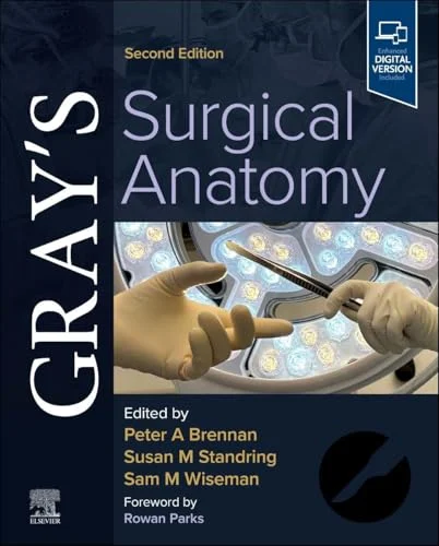
Gray’s Surgical Anatomy, 2nd edition (Videos Only)


Original price was: $187.25.$30.00Current price is: $30.00.
This Product is shared via google drive download link, So please share your correct Gmail id while placing the order .Please note that there are no CME points or certificate associated with this course Samples for Courses Can be found here : Free Samples Here!
Gray’s Surgical Anatomy, 2nd edition (Videos Only)
Gray’s Surgical Anatomy, 2nd Edition (Videos Only) is a specialized surgical anatomy video resource featuring cadaveric dissections, intraoperative demonstrations, anatomical approaches, and modern surgical techniques across major regions of the human body.
Overview
Gray’s Surgical Anatomy, 2nd Edition (Videos Only) provides a visual learning resource based on the supplementary surgical and anatomical videos associated with the second edition of Gray’s Surgical Anatomy, edited by Peter A. Brennan, Susan Standring, and Sam M. Wiseman.
Published by Elsevier, this updated edition combines detailed regional anatomy with contemporary operative approaches, emphasizing the anatomical structures, relationships, and technical considerations essential for safe surgical practice.
The accompanying video collection illustrates anatomical dissections and clinical procedures in head and neck surgery, neurosurgery, cardiothoracic surgery, abdominal surgery, urology, gynecology, orthopedic surgery, and other surgical specialties.
Particular emphasis is placed on the relationship between anatomical knowledge and operative technique, including robotic surgery, minimally invasive surgery, endoscopic procedures, and complex surgical dissections.
These videos are especially relevant for surgical residents, practicing surgeons, anatomy educators, and medical professionals seeking to strengthen their understanding of applied surgical anatomy.
Important: This is a videos-only educational product. The complete printed textbook, eBook, and PDF chapters are not included in this edition of the product.
Product Details
- Title: Gray’s Surgical Anatomy, 2nd edition (Videos Only)
- Edition: 2nd Edition
- Editors: Peter A. Brennan, Susan Standring, Sam M. Wiseman
- Publisher: Elsevier
- Publication Year: 2026
- Copyright Year: 2027
- ISBN of Reference Textbook: 9780443125683
- Format: Surgical Anatomy Videos
- Content Type: Cadaveric Dissections, Intraoperative Demonstrations, Robotic Surgery, Endoscopic and Minimally Invasive Procedures
- Primary Specialty: Surgery
- Related Specialties: Anatomy, Neurosurgery, Cardiothoracic Surgery, General Surgery, Urology, Orthopedics, Head and Neck Surgery
- Language: English
Target Audience
The Gray’s Surgical Anatomy, 2nd Edition video collection is suitable for:
- General Surgeons
- Surgical Residents and Fellows
- Medical Students
- Neurosurgeons
- Cardiothoracic Surgeons
- Head and Neck Surgeons
- Oral and Maxillofacial Surgeons
- Vascular Surgeons
- Orthopedic Surgeons
- Urologists
- Obstetricians and Gynecologists
- Radiologists
- Anatomists and Anatomy Educators
- Surgeons Preparing for Postgraduate Examinations
Program Overview
The educational videos complement the regional organization of Gray’s Surgical Anatomy, providing visual demonstrations of surgical dissections and operative anatomy.
1. Head and Neck Surgical Anatomy
Explore the intricate anatomy of the facial nerve, trigeminal nerve, orbit, infratemporal fossa, pterygopalatine fossa, pharynx, and neck.
The demonstrations include complex head and neck dissections, surgical access procedures, and selected robotic techniques.
2. Neurosurgical Anatomy
Study selected operative approaches involving intracranial structures, the ventricular system, skull base, and functional neurosurgery.
3. Spine and Regional Anatomy
Review anatomical relationships relevant to the back, lumbar puncture, epidural procedures, and spinal interventions.
4. Cardiothoracic Surgical Anatomy
Examine thoracic structures, mediastinal anatomy, cardiac access, the aortic arch, and selected complex cardiothoracic procedures.
5. Abdominal and Gastrointestinal Surgical Anatomy
Explore abdominal dissections, laparoscopic procedures, hepatobiliary surgery, liver transplantation, and gastrointestinal endoscopy.
6. Pelvic and Urogenital Surgical Anatomy
Review renal vascular anatomy, pelvic dissections, robotic prostatectomy, laparoscopic gynecologic procedures, and selected urological interventions.
7. Musculoskeletal and Lower Limb Anatomy
Study anatomical dissections of major joints, muscles, nerves, vascular structures, and clinically important lower limb regions.
Learning Objectives
After studying the relevant video demonstrations, learners should be better able to:
- Identify major anatomical structures and relationships encountered during surgical procedures.
- Recognize clinically important anatomical landmarks in cadaveric and intraoperative dissections.
- Relate regional anatomy to safe and effective operative approaches.
- Understand anatomical considerations in open, laparoscopic, robotic, and endoscopic surgery.
- Recognize structures at risk during complex dissections.
- Apply knowledge of surgical anatomy to operative planning and postgraduate surgical education.
- Compare selected conventional and minimally invasive approaches to surgical anatomy.
- Understand the importance of anatomical variation, procedural precision, and patient safety.
These are educational objectives derived from the video topics, not a separately published formal objectives list.
Official Publisher Video Topics
The following titles are drawn from Elsevier’s official supplementary video listing for the second edition. They describe the publisher’s video collection; the exact files included in this videos-only product should be checked against the available download.
Section 1 — Head and Neck
- 7.1 Facial nerve, dissection of left face
- 7.2 Trigeminal nerve, dissection of left face
- 7.3 Facial muscles, dissection of right face
- 8.1 Dissection of right orbit
- 10.1 Head and neck – hemisection
- 11.1 Head and neck – infratemporal fossa – superficial
- 11.2 Head and neck – infratemporal fossa – deep
- 11.3 Mandibular swing to access infratemporal fossa/maxillectomy
- 11.4 Total conservative parotidectomy with masticator and infratemporal fossa compartment resection
- 11.5 Double mandibular osteotomy for deep lobe parotid tumour/high internal carotid artery
- 11.6 Endoscopic anatomy of the infratemporal and pterygopalatine fossae
- 17.1 The neck
- 18.1 Head and neck – posterior – pharynx
- 18.2 Transoral robotic resection of right base of tongue tumour
- 19.1 Mucosal wave – stroboscopic examination of vocal folds
Section 2 — Neuroanatomy
- 22.1 Tuberculum sellae meningioma
- 22.2 Craniopharyngioma
- 24.1 Far-lateral approach to a right pontine cavernoma
- 25.1 Endoscopic third ventriculostomy
- 28.1 MR-guided focused ultrasound (MRgFUS) thalamotomy for essential tremor
Section 3 — Back and Spine
- 31.1 Back – superficial
- 36.1 Anatomy of lumbar puncture and epidural analgesia
- 36.2 Anatomy of lumbar puncture and epidural analgesia: lumbar puncture needle
Section 4 — Upper Limb
- 41.1 Upper limb – elbow joint
Section 5 — Thorax and Cardiothoracic Anatomy
- 43.1 Thorax – superficial
- 43.2 Chest wall resection
- 44.1 Thorax – heart
- 44.2 Thorax – internal
- 44.3 Replacement of aortic arch and placement of a frozen elephant trunk
- 44.4 Total aortic replacement
- 45.1 Dissection of the posterior and superior mediastinum
- 54.1 Thorax – breast
Section 6 — Abdomen and Gastrointestinal Surgery
- 56.1 Abdominal cavity part I
- 56.2 Abdominal cavity part II
- 58.1 Posterior view of female inguinal anatomy during robotic TAPP femoral hernia repair
- 59.1 Abdominal cavity part III
- 59.2 Abdominal cavity part IV
- 63.1 Laparascopic appendicectomy
- 65.1 Liver biopsy
- 65.2 Liver transplant
- 68.1 Laparoscopic adrenalectomy for phaeochromocytoma in patients with MEN 2A
- 70.1 Barrett’s oesophagus
- 70.2 Dysplastic Barrett’s – endoscopic mucosal resection
- 70.3 ERCP with cholangioscopy
- 70.4 Ascending colon polyp – endoscopic mucosal resection
- 70.5 Rectal lesion – endoscopic submucosal dissection
Section 7 — Pelvic and Urogenital Surgery
- 72.1 Using 3D perfusion zone models during robotassisted partial nephrectomy
- 72.2 Identifying renal vascular anatomy using intravenous indocyanine green during robotic partial nephrectomy
- 73.1 Pelvis and perineum – female
- 73.2 Pelvis and perineum – male
- 74.1 Robotic pelvic lymph node dissection
- 76.1 Transurethral resection of the prostate (TURP)
- 76.2 Holmium enucleation of the prostate (HoLEP)
- 76.3 Robotic prostatectomy – interfascial nerve spare
- 76.4 Robotic prostatectomy – urethrovesical anastomosis
- 77.1 Total laparoscopic hysterectomy with right salpingo-oophorectomy
- 77.2 Laparoscopic right salpingo-oophorectomy
- 77.3 Robotic dissection of inferior hypogastric nerve on the left side
Section 8 — Bony Pelvis and Lower Limb
- 81.1 Lower limb – thigh and leg – posterior – deep
- 82.1 Lower limb – gluteal region – superficial
- 82.2 Lower limb – thigh – anterior – deep
- 82.3 Lower limb – leg and foot – anterior – deep
- 83.1 Lower limb – gluteal region – deep
- 84.1 Cadaveric dissection of the right hip joint
- 85.1 Superficial dissection of the left femoral triangle, female
- 85.2 Superficial dissection of lower limb, posterior aspect
- 87.1 Lower limb – knee joint
- 89.1 Lower limb – ankle joint
- 90.1 Lower limb – foot – plantar
- 90.2 Lower limb – leg and foot – anterior – superficial
Major Topics Covered
- Applied Surgical Anatomy
- Cadaveric Dissection Videos
- Intraoperative Surgical Anatomy
- Head and Neck Anatomy
- Cranial Nerve Dissection
- Neurosurgical Anatomy
- Skull Base Surgical Approaches
- Cardiothoracic Surgical Anatomy
- Aortic Surgery
- Abdominal Surgical Anatomy
- Laparoscopic Surgery
- Liver Transplantation
- Robotic Surgery
- Endoscopic Anatomy
- Pelvic and Urogenital Anatomy
- Robotic Prostatectomy
- Minimally Invasive Gynecologic Surgery
- Upper and Lower Limb Anatomy
- Surgical Landmarks and Anatomical Hazards
- Surgical Training and Anatomy Education
Editors
Peter A. Brennan, OBE, MD, PhD, FRCS, FRCSI, Hon FRCSGlas, FRCSEd
Consultant Maxillofacial and Head and Neck Surgeon and Honorary Professor of Surgery, Queen Alexandra Hospital, Portsmouth, United Kingdom.
Susan Standring, MBE, PhD, DSc, Hon FAS, Hon FRCS
Emeritus Professor of Anatomy, King’s College London, United Kingdom, and Editor-in-Chief of Gray’s Anatomy.
Sam M. Wiseman, BSc, MD, FRCSC, FACS
Professor of Surgery, University of British Columbia, Canada, specializing in general surgery, surgical oncology, and head and neck surgery.
Why This Video Collection Is Relevant
A thorough understanding of regional and applied anatomy is fundamental to safe surgical practice.
The Gray’s Surgical Anatomy, 2nd Edition video resource offers opportunities to observe anatomical structures in cadaveric dissections and operative environments rather than relying entirely on static illustrations.
Key Educational Benefits
- Visualize clinically important anatomical structures and surgical relationships.
- Review real-world operative approaches in multiple surgical disciplines.
- Strengthen anatomical knowledge for surgical training and examinations.
- Explore minimally invasive, laparoscopic, robotic, and endoscopic techniques.
- Recognize anatomical structures that require particular attention during surgical procedures.
- Connect anatomical theory with practical operative applications.
- Support continuing surgical education and procedural anatomy revision.
This video resource is particularly useful for surgical trainees and practicing surgeons seeking a more visual understanding of the anatomy underlying modern surgical procedures.
Short Description
Gray’s Surgical Anatomy, 2nd Edition (Videos Only) is a surgical anatomy video resource featuring cadaveric dissections, operative demonstrations, robotic surgery, endoscopic procedures, and region-specific surgical anatomy.
Edited by Peter A. Brennan, Susan Standring, and Sam M. Wiseman, this Elsevier resource supports surgical education and practical anatomical understanding across multiple surgical specialties.
Key Topics:
- Head and Neck Dissections
- Neurosurgical and Skull Base Anatomy
- Cardiothoracic Surgical Procedures
- Abdominal and Hepatobiliary Surgery
- Robotic and Laparoscopic Surgery
- Pelvic and Urogenital Anatomy
- Musculoskeletal and Lower Limb Dissections
Format: Videos Only — No Textbook or PDF Included.
Ideal for: Surgeons, Surgical Residents, Medical Students, Neurosurgeons, Cardiothoracic Surgeons, Anatomists, and Surgical Educators.
Related products
General Surgery
Cardiology
General Surgery
Critical Care - Emergency medicine
Critical Care - Emergency medicine
Need-to-Know Emergency Medicine: A Review for Physicians in a Hurry 2020

Obstetrics & Gynecology
2023 Surgical Pathology Update: Diagnostic Pearls for the Practicing Pathologist: Vol. VII