
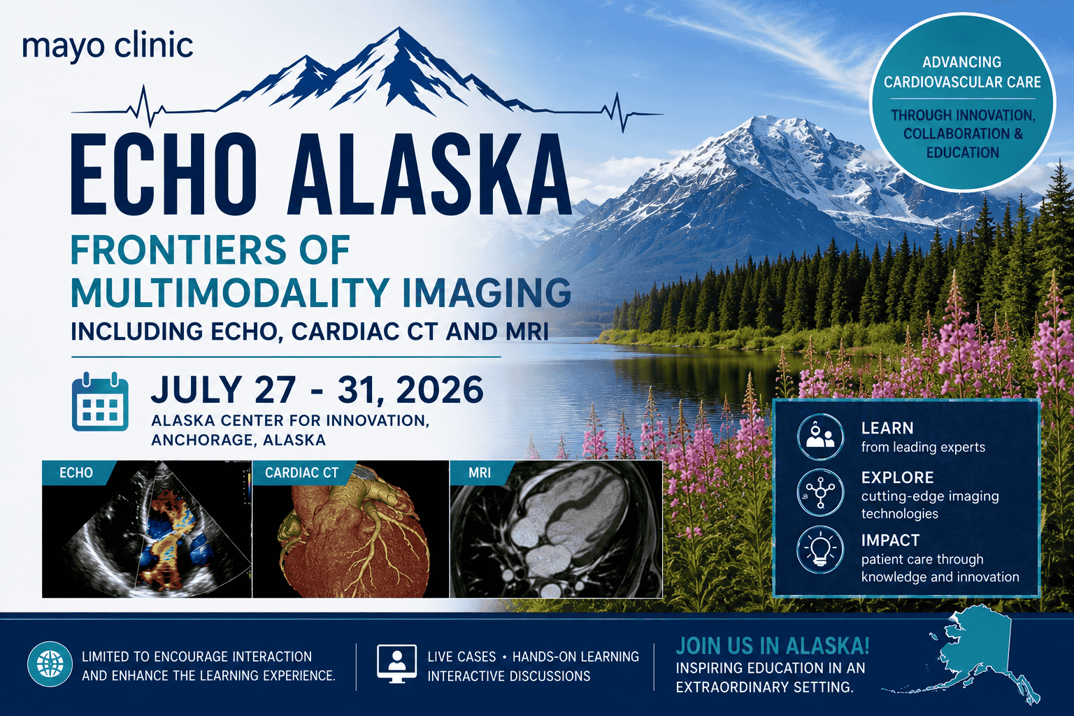
Mayo Clinic Echo Alaska Frontiers of Multimodality Imaging Including Echo, Cardiac CT and MRI July 27 – 31, 2026
$890.00 Original price was: $890.00.$300.00Current price is: $300.00.
This Product is shared via google drive download link, So please share your correct Gmail id while placing the order .Please note that there are no CME points or certificate associated with this course Samples for Courses Can be found here : Free Samples Here!
EXPRESS DISCOVER PayPal
Mayo Clinic Echo Alaska 2026 – Frontiers of Multimodality Imaging Including Echo, Cardiac CT and MRI
Echocardiography • Cardiac CT • Cardiac MRI • Nuclear Imaging • 3-D Echo
Develop a practical, integrated approach to modern cardiovascular imaging with Mayo Clinic Echo Alaska: Frontiers of Multimodality Imaging Including Echo, Cardiac CT and MRI 2026.
Held July 27–31, 2026, at the Marriott Anchorage Downtown in Anchorage, Alaska, this five-day Mayo Clinic cardiovascular imaging symposium provides an extensive review of contemporary:
- Echocardiography
- 2-D and 3-D echocardiography
- Cardiac CT
- Cardiac MRI
- Nuclear cardiology
- PET imaging
- PYP imaging
- Strain imaging
- Point-of-care ultrasound
- Procedural imaging
The course emphasizes not simply how each imaging modality works, but how to choose and integrate Echo, CT, MRI, and nuclear imaging for clinical decision-making.
Major disease areas include:
- Heart failure
- Cardiomyopathy
- Valvular heart disease
- Ischemic heart disease
- Congenital heart disease
- Cardiovascular disease in pregnancy
- Aortic disease
- Cardiac masses
- Unstable cardiovascular patients
- Structural and interventional heart disease
A unique component of the original program was live echocardiographic scanning, allowing faculty to demonstrate comprehensive image acquisition and illustrate how real-time echocardiographic findings influence patient management.
Course Details
- Course: Echo Alaska: Frontiers of Multimodality Imaging Including Echo, Cardiac CT and MRI
- Provider: Mayo Clinic
- Year: 2026
- Dates: July 27–31, 2026
- Duration: 5 Days
- Venue: Marriott Anchorage Downtown
- Location: Anchorage, Alaska, USA
- Primary Specialty: Cardiology / Cardiovascular Imaging
- Additional Specialty: Radiology / Echocardiography
- Modalities: Echo / Cardiac CT / Cardiac MRI / Nuclear Imaging
- Original Activity: 23.0 Credit Hours
- MedicalAmboss Delivery: Google Drive
- MedicalAmboss CME / Certificate: Not Included
- Language: English
Course Overview
Cardiovascular imaging increasingly requires clinicians to select among several complementary technologies rather than relying on a single test.
A patient with structural or cardiovascular disease may undergo:
Echocardiography → Cardiac CT → Cardiac MRI → Nuclear Imaging
depending on the clinical question.
Each modality provides different information.
The central principle of multimodality cardiovascular imaging is therefore:
Choose the Right Modality for the Right Patient and the Right Clinical Question
This Mayo Clinic course explores both the individual strengths of each imaging method and their incremental value when used together.
Learning Objectives
Upon completion of the original program, participants should be better able to:
- Utilize cardiovascular imaging to support clinical decision-making.
- Integrate echocardiography into the management of valvular heart disease.
- Interpret cardiovascular imaging in patients with heart failure.
- Use cardiac CT and MRI to complement echocardiographic findings.
- Apply multimodality imaging to congenital heart disease.
- Recognize the strengths and limitations of each cardiovascular imaging modality.
- Select CT, MRI, echo, or nuclear imaging according to the clinical question.
- Review contemporary imaging of cardiomyopathy.
- Apply imaging to ischemic heart disease.
- Review imaging in unstable and critically ill patients.
- Improve 2-D and 3-D echocardiographic acquisition.
- Use echocardiography for procedural and interventional guidance.
- Review advanced sonographic techniques including strain imaging.
- Improve recognition of cardiovascular pathology through case-based multimodality interpretation.
HEART FAILURE & MULTIMODALITY IMAGING
Introduction to Cardiac MRI & CT: Which Test for Which Patient?
One of the first major questions addressed by the program is:
Which cardiac imaging test should be ordered for which patient?
The choice depends on:
- Clinical indication
- Cardiac anatomy
- Ventricular function
- Tissue characterization needs
- Coronary assessment
- Valve disease
- Patient characteristics
- Previous imaging findings
Echocardiography
Echocardiography remains a foundational cardiovascular imaging technique.
It can provide information regarding:
- Ventricular size
- Ventricular function
- Valve morphology
- Valve hemodynamics
- Chamber size
- Pulmonary pressures
- Pericardial disease
- Congenital abnormalities
Its portability and real-time imaging also make it particularly useful in acute clinical settings.
Cardiac CT
Cardiac CT can provide detailed anatomical information involving:
- Coronary arteries
- Aorta
- Cardiac chambers
- Valve anatomy
- Calcification
- Structural heart anatomy
- Procedural planning
CTA is also incorporated into the course for:
- Chest pain
- Valvular heart disease
- Aortic disease
Cardiac MRI
Cardiac MRI offers powerful assessment of:
- Ventricular function
- Cardiac morphology
- Myocardial tissue
- Cardiomyopathy
- Inflammation
- Fibrosis
- Congenital heart disease
The value of MRI often lies in combining anatomy, function, and tissue characterization.
Multimodality Imaging in Cardiomyopathy
The course includes dedicated education on multimodality imaging in cardiomyopathy.
A patient with suspected cardiomyopathy may require integration of:
- Echocardiographic phenotype
- Ventricular function
- Cardiac MRI tissue characterization
- CT where clinically appropriate
- Nuclear imaging in selected conditions
The goal is to answer:
What Is the Cardiomyopathy, How Severe Is It, and Does Imaging Change Management?
Systolic & Diastolic Heart Failure
The program reviews imaging in both:
- Systolic dysfunction
- Diastolic dysfunction
Evaluation may involve:
- Ejection fraction
- Chamber size
- Filling patterns
- Hemodynamics
- Valve disease
- Pulmonary pressures
- Myocardial structure
Diastology
Diastolic function remains an important component of comprehensive echocardiographic examination.
Interpretation can integrate:
- Mitral inflow
- Tissue Doppler
- Left atrial size
- Pulmonary pressures
- Other echocardiographic parameters
The original course also includes practical live scanning designed to reinforce image acquisition and interpretation.
VALVULAR HEART DISEASE
CTA for the Evaluation of Valvular Heart Disease
Cardiac CT has an expanding role in structural and valvular heart disease.
Potential contributions include:
- Valve anatomy
- Calcification assessment
- Aortic anatomy
- Procedural planning
- Structural relationships
Echocardiography in Valve Disease
Echo remains central to evaluating:
- Aortic stenosis
- Aortic regurgitation
- Mitral stenosis
- Mitral regurgitation
- Tricuspid valve disease
- Other structural abnormalities
A comprehensive evaluation involves both:
Anatomy + Hemodynamics
2-D & 3-D Echocardiography
An important focus of Echo Alaska is the use of 2-D and 3-D echocardiography.
3-D imaging can enhance visualization of:
- Valve morphology
- Leaflet anatomy
- Structural relationships
- Interventional targets
This is particularly relevant in modern structural heart interventions.
Imaging for Interventional Guidance
The course reviews how echocardiography can support cardiovascular interventions through:
- Preprocedural assessment
- Intraprocedural imaging
- Device positioning
- Anatomical visualization
- Assessment of procedural results
- Recognition of complications
3-D Image Acquisition
The practical workshop addresses:
- Acquisition
- Knobology
- Optimization
- Intraoperative imaging
- Procedural imaging
- Troubleshooting
The objective is to improve the quality and clinical usefulness of 3-D datasets.
ISCHEMIC HEART DISEASE
The program includes a dedicated section on:
Ischemic Heart Disease and Assessment of the Unstable Patient
with several complementary imaging approaches.
CTA for Chest Pain
Coronary CTA can be useful in selected patients presenting with chest pain.
The course reviews how CT may contribute to:
- Coronary assessment
- Anatomical evaluation
- Clinical decision-making
Nuclear Perfusion Imaging for Ischemia
The program includes dedicated education in nuclear perfusion imaging.
Nuclear techniques can support assessment of:
- Myocardial perfusion
- Ischemia
- Functional significance
- Selected cardiomyopathies
Stress Echocardiography
Stress Echo for Ischemia and Beyond reviews the application of stress echocardiography to ischemic evaluation as well as broader clinical questions.
Stress echocardiography combines:
- Physiologic stress
- Wall-motion assessment
- Hemodynamic information
- Real-time cardiac imaging
Nuclear Cardiology
Although the official course title emphasizes Echo, CT, and MRI, the 2026 curriculum also includes nuclear imaging.
Topics include:
- Nuclear perfusion
- PET
- PYP imaging
- Image acquisition
- Interpretation
- Troubleshooting
- Interesting cases
PET & PYP Imaging
The nuclear workshop includes:
PET, PYP, and More
providing practical discussion of nuclear cardiovascular imaging and its use in selected clinical scenarios.
POCUS & ECHO IN THE ICU
The curriculum includes:
POCUS & Echo in the ICU
highlighting cardiovascular ultrasound in unstable and critically ill patients.
Point-of-Care Ultrasound
POCUS can provide rapid bedside information about:
- Cardiac function
- Volume status
- Pericardial fluid
- Lung findings
- Hemodynamic status
It is particularly useful when rapid clinical decisions are required.
Imaging the Unstable Patient
In acute illness, imaging must answer focused questions efficiently.
A practical approach is:
What Is the Immediate Clinical Question? → Which Modality Can Answer It Quickly and Safely?
Bedside ultrasound can therefore complement more comprehensive imaging when immediate information is required.
CONGENITAL HEART DISEASE & PREGNANCY
The course includes multiple case-based sessions addressing:
- Adult congenital heart disease
- Pediatric/congenital cardiovascular anatomy
- Pregnancy-related cardiovascular imaging
Multimodality Imaging in Congenital Heart Disease
Congenital heart disease often requires integration of:
- Echocardiography
- CT
- MRI
because complex anatomy may not be completely defined by a single modality.
Congenital Cardiac Imaging
Important questions may include:
- What is the underlying anatomy?
- What prior intervention has been performed?
- What is the ventricular function?
- Are there residual shunts or obstructions?
- Is additional intervention required?
Pregnancy & Cardiovascular Imaging
Cardiac disease during pregnancy requires special attention to:
- Maternal physiology
- Fetal considerations
- Radiation exposure
- Contrast use
- Appropriate test selection
Imaging choices therefore require careful risk-benefit assessment.
MULTIMODALITY IMAGING WORKSHOPS
One of the strongest components of Echo Alaska is its practical workshop structure.
The program includes:
- CT/MRI workshop
- Nuclear imaging workshop
- Sonography workshop
- Procedural echo workshop
- Live echo scanning
- Strain workshop
- 3-D valve imaging
- Congenital imaging demonstrations
CT/MRI Workshop
The CT/MRI workshop focuses on:
- Troubleshooting
- Image acquisition
- Interpretation
- Interesting clinical cases
This helps move the curriculum beyond basic modality descriptions into practical image interpretation.
Tips & Tricks for Sonographers
The course includes a focused sonography session covering practical imaging techniques.
Topics include:
- Coronary imaging
- POCUS
- Lung ultrasound
- Pedoff probe use
- Other acquisition tips
Comprehensive Echocardiographic Acquisition
High-quality interpretation depends on high-quality image acquisition.
The course emphasizes that important diagnostic findings can be missed when the examination is incomplete.
A structured echo study should therefore be:
Comprehensive + Reproducible + Clinically Focused
ECHO LIVE SCANNING
A defining component of the original Echo Alaska course is live patient scanning.
This allows participants to observe:
- Probe positioning
- Image optimization
- Real-time decision-making
- Acquisition technique
- Interpretation
Live Strain Workshop
The program includes live demonstrations focused on strain imaging.
Strain may provide additional information about myocardial deformation beyond conventional ejection fraction.
Strain Imaging
Potential applications can include selected evaluation of:
- Cardiomyopathy
- Heart failure
- Subclinical ventricular dysfunction
- Other myocardial disease
Interpretation should always be integrated with the broader clinical and echocardiographic context.
Live 3-D & Valve Imaging
Another live demonstration focuses on:
- 3-D echocardiography
- Valve imaging
The session provides practical exposure to how high-quality 3-D data are acquired rather than only showing final reconstructed images.
Live Congenital Imaging
Congenital and other cardiovascular cases are also included in the live scanning curriculum.
This reinforces the importance of adapting image acquisition to the patient’s underlying anatomy.
AORTIC DISEASE
The final-day curriculum includes:
Multimodality Imaging for Aortic Disease
Imaging the Aorta
Different modalities may contribute to evaluation of:
- Aortic dimensions
- Aortic valve relationships
- Aortic wall disease
- Acute aortic syndromes
- Surveillance
CT and MRI can be particularly valuable for comprehensive aortic anatomical assessment.
CARDIAC MASSES
The program includes:
Cardiac Masses: Imaging/Pathologic Correlation
This topic illustrates the value of combining:
- Echocardiography
- CT
- MRI
- Pathologic correlation
when evaluating an intracardiac mass.
Imaging a Cardiac Mass
A practical assessment may consider:
- Location
- Size
- Attachment
- Mobility
- Tissue characteristics
- Hemodynamic effect
The differential may include:
- Tumor
- Thrombus
- Vegetation
- Other structural abnormalities
No single modality necessarily answers every question.
Case-Based Learning
A major feature of the 2026 program is extensive case-based learning.
Rather than presenting only conventional lectures, the curriculum repeatedly uses clinical cases to ask:
What Imaging Would You Order? → What Does It Show? → What Additional Modality Is Needed? → Does It Change Management?
This approach closely reflects real-world cardiovascular practice.
Strengths & Limitations of Imaging Modalities
Echocardiography
Strengths
- Real-time
- Portable
- Hemodynamic information
- No ionizing radiation
Limitations
- Image quality can vary
- Operator dependent
- Some anatomical regions may be difficult to visualize
Cardiac CT
Strengths
- Excellent spatial resolution
- Coronary anatomy
- Aortic imaging
- Structural planning
Limitations
- Ionizing radiation
- Contrast considerations
- Limited tissue characterization compared with MRI
Cardiac MRI
Strengths
- Excellent ventricular assessment
- Tissue characterization
- No ionizing radiation
- Congenital imaging
Limitations
- Longer examination
- Device / patient compatibility considerations
- Availability
- Patient tolerance
Nuclear Imaging
Strengths
- Physiologic / perfusion information
- Disease-specific applications
- PET and molecular imaging potential
Limitations
- Radiation exposure
- Availability
- Lower anatomical resolution compared with CT/MRI in some applications
Major Topics Covered
- Echocardiography
- Cardiovascular Imaging
- Multimodality Imaging
- Cardiac CT
- Cardiac CTA
- Cardiac MRI
- CMR
- Nuclear Cardiology
- PET Imaging
- PYP Imaging
- 2-D Echocardiography
- 3-D Echocardiography
- Strain Imaging
- POCUS
- Lung Ultrasound
- Heart Failure
- Diastology
- Cardiomyopathy
- Valvular Heart Disease
- Structural Heart Disease
- Ischemic Heart Disease
- Stress Echocardiography
- Coronary CTA
- Congenital Heart Disease
- Cardiovascular Disease in Pregnancy
- Interventional Echocardiography
- Procedural Imaging
- Aortic Disease
- Cardiac Masses
- Sonography
- Live Echocardiographic Scanning
Who Should Take This Course?
The official Mayo Clinic audience includes:
- Adult Cardiovascular Specialists
- Congenital Cardiovascular Specialists
- Cardiologists
- Internists
- Anesthesiologists
- Critical Care Physicians
- Cardiovascular Surgeons
- Other Surgeons involved in cardiovascular care
- Sonographers
The material is also highly relevant to:
- Cardiac Imaging Specialists
- Echocardiographers
- Cardiac Radiologists
- Cardiovascular Fellows
- Cardiology Fellows
- Echocardiography Technologists
- Structural Heart Teams
Why This Course Is Useful
The principal strength of Mayo Clinic Echo Alaska 2026 is its integrated approach.
Instead of treating each imaging technique as an isolated specialty, the program asks how modalities complement one another.
Learners can review:
- Echo vs. CT vs. MRI selection
- Heart failure imaging
- Cardiomyopathy
- Valvular heart disease
- Cardiac CTA
- Ischemia imaging
- Nuclear perfusion
- Stress echo
- POCUS
- Adult congenital heart disease
- Pregnancy
- PET/PYP
- Image acquisition
- 3-D echocardiography
- Strain
- Interventional guidance
- Aortic disease
- Cardiac masses
The overall learning pathway can be summarized as:
Clinical Question → Echo → CT / MRI / Nuclear When Needed → Integrated Interpretation → Management Decision
Original Mayo Clinic Accreditation
The original Mayo Clinic Echo Alaska 2026 activity offered up to:
- 23.0 AMA PRA Category 1 Credits™
- 23.0 ABIM MOC Points
- 23.0 ABS MOC Points
- 23.0 ANCC Credits
for eligible participants completing the official Mayo Clinic activity requirements.
Important MedicalAmboss CME Notice
The MedicalAmboss digital product does not include CME points or a certificate.
Therefore, do not advertise this product as:
- 23 CME Credits Included
- 23 ABIM MOC Included
- 23 ABS MOC Included
- 23 ANCC Credits Included
- Official Mayo Clinic CME Certificate Included
unless the purchaser is actually receiving official Mayo Clinic registration and credit eligibility.
The appropriate wording is:
Original Mayo Clinic Activity: Up to 23.0 Credit Hours
and separately:
MedicalAmboss Product: CME / Certificate Not Included
Product / Delivery Information
- Product: Mayo Clinic Echo Alaska: Frontiers of Multimodality Imaging Including Echo, Cardiac CT and MRI 2026
- Provider: Mayo Clinic
- Dates: July 27–31, 2026
- Location: Anchorage, Alaska
- Venue: Marriott Anchorage Downtown
- Specialty: Cardiology / Cardiovascular Imaging
- Modalities: Echo / CT / MRI / Nuclear
- Original Activity: 23.0 Credit Hours
- Delivery: Google Drive
- CME Points with MedicalAmboss: Not Included
- Certificate: Not Included
4. Short Description
Mayo Clinic Echo Alaska: Frontiers of Multimodality Imaging Including Echo, Cardiac CT and MRI 2026 provides an advanced five-day review of modern cardiovascular multimodality imaging.
Topics include echocardiography, cardiac CT, cardiac MRI, nuclear perfusion imaging, PET/PYP, heart failure, cardiomyopathy, valvular heart disease, ischemic heart disease, POCUS, congenital heart disease, pregnancy, 2-D and 3-D echo, strain imaging, procedural guidance, aortic disease, and cardiac masses.
The original course also features CT/MRI and nuclear workshops, tips and tricks for sonographers, and live echocardiographic scanning demonstrations.
+ Topics:
Monday, July 27, 2026
-
6:30 a.m.: Precourse Registration and Continental Breakfast
-
7:00 a.m.: Welcome and Introduction – Course Overview & Objective Review (Grace Lin, MD, MBA)
HEART FAILURE AND INTRODUCTION TO MULTIMODALITY IMAGING (Moderator: Grace Lin, MD, MBA)
-
7:10 a.m.: Introduction to Cardiac MRI & CT: Which Test for Which Patient (Suhny Abbara, MD)
-
7:40 a.m.: Case 1 (Grace Lin, MD, MBA)
-
8:00 a.m.: Case 2 (Daniel Borgeson, MD)
-
8:20 a.m.: Case 3 (Grace Lin, MD, MBA)
-
8:40 a.m.: Case 4 (S. Allen Luis, MBBS, PhD)
-
9:00 a.m.: Questions With Panel Including Surgeon
-
9:20 a.m.: Refreshment Break & Visit Exhibits
SYSTOLIC AND DIASTOLIC HEART FAILURE: ECHO/CT/MRI (Moderator: Grace Lin, MD, MBA)
-
9:40 a.m.: Multimodality Imaging in Cardiomyopathy (John Bois, MD)
-
10:10 a.m.: Case 6 (Daniel Borgeson, MD)
-
10:30 a.m.: Case 7 (Panithaya Chareonthaitawee, MD)
-
10:50 a.m.: Case 8 (Matthew Martinez, MD)
-
11:10 a.m.: Case 9 (Grace Lin, MD, MBA)
-
11:30 a.m.: Case 10 (Daniel Borgeson, MD)
-
11:50 a.m.: Questions With Panel Including Surgeon
-
12:15 p.m.: Grab lunch and return to ballroom for product theater, and then return for evening session.
-
12:45 p.m.: Non-CME Product Theater – Sponsored by AstraZeneca – No CME Credit
-
1:45 p.m.: Adjourn until 4 p.m.
Tuesday, July 28, 2026
-
6:30 a.m.: Continental Breakfast – Visit Exhibits
VALVULAR HEART DISEASE I
-
7:00 a.m.: CTA for the Evaluation of Valvular Heart Disease (Suhny Abbara, MD)
-
7:30 a.m.: Case 1 (Vuyisile Nkomo, MD, MPH)
-
7:50 a.m.: Case 2 (Matthew Martinez, MD)
-
8:10 a.m.: Case 3 (Jared Bird, MD)
-
8:30 a.m.: Case 4 (S. Allen Luis, MBBS, PhD)
-
8:50 a.m.: Questions With Panel Including Surgeon
-
9:10 a.m.: Refreshment Break & Visit Exhibits
VALVULAR HEART DISEASE II
-
9:40 a.m.: Case 5 (Vuyisile Nkomo, MD, MPH)
-
10:00 a.m.: Case 6 (Rekha Mankad, MD)
-
10:20 a.m.: Case 7 (Alberto Pochettino, MD)
-
10:40 a.m.: Case 8 (Rekha Mankad, MD)
-
11:00 a.m.: Case 9 (Nathaniel Taggart, MD)
-
11:20 a.m.: Case 10 (S. Allen Luis, MBBS, PhD)
-
11:40 a.m.: Questions With Panel Including Surgeon
-
12:00 p.m.: Adjourn. Grab lunch and return to ballroom for product theater.
-
12:30 p.m.: Non-CME Product Theater Sponsored by Kiniksa – No CME Credit
Wednesday, July 29, 2026
-
6:30 a.m.: Continental Breakfast – Visit Exhibits
ISCHEMIC HEART DISEASE AND ASSESSMENT OF THE UNSTABLE PATIENT (Moderator: Crystal Bonnichsen, MD)
-
12:00 a.m.: POCUS & Echo in the ICU (Jared Bird, MD)
-
7:00 a.m.: CTA for Chest Pain (Philip Araoz, MD)
-
7:25 a.m.: Nuclear Perfusion Imaging for Evaluation of Ischemia (Panithaya Chareonthaitawee, MD)
-
7:50 a.m.: Stress Echo for Ischemia and Beyond (S. Allen Luis, MBBS, PhD)
-
8:40 a.m.: Questions With Panel Including Surgeon
-
9:00 a.m.: Refreshment Break & Visit Exhibits
CONGENITAL HEART DISEASE AND PREGNANCY (Moderator: Jonathan Johnson, MD)
-
9:30 a.m.: Case 1 (Steve Kindel, MD)
-
9:50 a.m.: Case 2 (M. Yasir Qureshi, MBBS)
-
10:10 a.m.: Case 3 (Nathaniel Taggart, MD)
-
10:30 a.m.: Case 4 (Jonathan Johnson, MD)
-
10:50 a.m.: Case 5 (M. Yasir Qureshi, MBBS)
-
11:10 a.m.: Case 6 (Steve Kindel, MD)
-
11:30 a.m.: Questions With Panel Including Surgeon
-
12:00 p.m.: Adjourn
Thursday, July 30, 2026
-
6:30 a.m.: Continental Breakfast – Visit Exhibits
MULTIMODALITY IMAGING WORKSHOPS & ECHO LIVE SCANNING
-
12:00 a.m.: CT/MRI Workshop – Trouble Shooting, Image Acquisition, Interpretation, Interesting Cases (Ballroom 2) (Suhny Abbara, MD, Philip Araoz, MD, John Bois, MD)
-
12:00 a.m.: Nuclear Workshop – PET, PYP, and More – Trouble Shooting, Image Acquisition, Interpretation, Interesting Cases (Ballroom 2) (John Bois, MD, Panithaya Chareonthaitawee, MD)
-
7:00 a.m.: Tips and Tricks for the Sonographer (including hands-on): Imaging Tips (Coronaries, POCUS/Lung US, Pedoff Probe, etc.) (Main Ballroom) (Ahmed Sami Abuzaid, MD, Macy Bartosh, RDCS, Jared Bird, MD, Crystal Bonnichsen, MD, Nicholas Enger, RDCS, Donald Hagler, MD, Steve Kindel, MD, M. Yasir Qureshi, MBBS)
-
8:00 a.m.: Echo for Guiding Procedures: 3-D Image Acquisition; focus on knobology and optimization, tips and tricks for intraop and procedural imaging (Main Ballroom) (Macy Bartosh, RDCS, Jared Bird, MD, Nicholas Enger, RDCS, Donald Hagler, MD, Rekha Mankad, MD, Vuyisile Nkomo, MD, MPH, Nathaniel Taggart, MD)
-
9:30 a.m.: Refreshment Break & Visit Exhibits
ECHO LIVE SCANNING (Main Ballroom)
-
10:00 a.m.: Live Case Demonstrations – Strain Workshop (Ahmed Sami Abuzaid, MD, Macy Bartosh, RDCS, Daniel Borgeson, MD, Nicholas Enger, RDCS, Grace Lin, MD, MBA, Matthew Martinez, MD)
-
10:40 a.m.: Live Case Demonstrations – 3-D and Valves (Ahmed Sami Abuzaid, MD, Macy Bartosh, RDCS, Jared Bird, MD, Nicholas Enger, RDCS, Vuyisile Nkomo, MD, MPH)
-
11:20 a.m.: Live Case Demonstrations – Congenital/Other (Ahmed Sami Abuzaid, MD, Nicholas Enger, RDCS, Donald Hagler, MD, Jonathan Johnson, MD, Steve Kindel, MD, M. Yasir Qureshi, MBBS, Nathaniel Taggart, MD)
-
12:00 p.m.: Adjourn
Friday, July 31, 2026
-
6:30 a.m.: Continental Breakfast – Visit Exhibits
MISCELLANEOUS TOPICS (Moderator: Crystal Bonnichsen, MD)
-
12:00 a.m.: Case of the Year – You Vote! (Crystal Bonnichsen, MD)
-
7:00 a.m.: Multimodality Imaging for Aortic Disease (Crystal Bonnichsen, MD)
-
7:25 a.m.: Cardiac Masses: Imaging/Pathologic Correlation (Philip Araoz, MD, Melanie Bois, MD)
-
8:15 a.m.: Refreshment Break
-
10:00 a.m.: Adjourn
| Select | Full course Videos+Qbank, Only Videos, Only Qbank |
|---|
Related products

Medical Accounts
Cardiology
Internal Medicine
Internal Medicine
Critical Care - Emergency medicine

USMLE / kaplan and Board Review
Uworld USMLE COMLEX Level 1 Qbank, Updated Jan 2023, System- and Subject-wise (PDF)
Cardiology
Critical Care - Emergency medicine
2022 UPDATES IN THERAPEUTICS: CRITICAL CARE PHARMACY PREPARATORY REVIEW AND RECERTIFICATION COURSE