
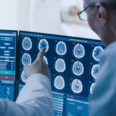
Oakstone UCSF Radiology Review Comprehensive Imaging 2026


$35.00
This Product is shared via google drive download link, So please share your correct Gmail id while placing the order .Please note that there are no CME points or certificate associated with this course Samples for Courses Can be found here : Free Samples Here!
Oakstone UCSF Radiology Review Comprehensive Imaging 2026
Diagnostic Radiology • Comprehensive Imaging • Board Review • Case-Based Radiology
Original Release: August 16, 2026 | 35 Hours | 60+ Video Sessions
Strengthen image interpretation, differential diagnosis, imaging utilization, protocol selection, and board preparation with Oakstone UCSF Radiology Review Comprehensive Imaging 2026.
Officially released by Oakstone as UCSF Radiology: Comprehensive Review, this comprehensive online video program delivers a practical, case-driven review of diagnostic radiology across all major organ systems and imaging modalities.
Developed with faculty from the University of California, San Francisco (UCSF) Department of Radiology and Biomedical Imaging, the program includes 60+ in-depth sessions combining:
- Didactic radiology lectures
- Rapid-fire imaging cases
- CORE-style case review
- Differential diagnosis
- Imaging protocol selection
- Board-focused teaching
- Imaging safety
- Nuclear medicine
- Interventional radiology
- Non-interpretive skills
The official activity requires approximately 35 hours to complete and was designed both for radiologists preparing for certification examinations and clinicians seeking a broad review of contemporary imaging practice.
Course Details
- Product: Oakstone UCSF Radiology Review Comprehensive Imaging 2026
- Official Title: UCSF Radiology: Comprehensive Review
- Provider: Oakstone / CMEinfo
- Accrediting Institution: University of California, San Francisco – UCSF
- Year: 2026
- Original Release Date: August 16, 2026
- Credit Deadline: August 15, 2029
- Estimated Time to Complete: 35.00 Hours
- Number of Sessions: 60+
- Format: Online Video
- Primary Specialty: Radiology
- Educational Style: Case-Based + Didactic + Rapid-Fire Review
- Course Chair: Soonmee Cha, MD
- Primary Focus: Comprehensive Diagnostic Imaging and Differential Diagnosis
- Official CME: Up to 35.00 AMA PRA Category 1 Credits™
The official curriculum includes major teaching blocks covering:
- Abdomen & Pelvis
- GI / GU
- Thoracic Imaging
- Breast Imaging
- Pediatric Radiology
- Neuroradiology
- Neurovascular Imaging
- Musculoskeletal Radiology
- Obstetric & Gynecologic Ultrasound
- Nuclear Medicine
- Interventional Radiology
- Radiation Safety
- Quality Assurance
- Non-Interpretive Skills
Course Chair
Soonmee Cha, MD
- Professor of Radiology
- Vice-Chair for Education
- Director, Radiology Residency
- University of California, San Francisco
Dr. Cha leads a multidisciplinary UCSF faculty representing abdominal imaging, thoracic imaging, neuroradiology, musculoskeletal imaging, breast imaging, pediatrics, nuclear medicine, and interventional radiology.
Intended Audience
The official intended audience includes:
- Radiologists
- Physicians
- Other medical professionals who would benefit from a greater understanding of imaging protocols and differential diagnosis
This course is particularly useful for:
- Diagnostic Radiologists
- Radiology Residents
- Radiology Fellows
- Academic Radiologists
- Community Radiologists
- Physicians Preparing for Radiology Board Examinations
- Imaging Specialists
- Physicians Seeking Multimodality Imaging Review
The strongest audience remains:
Diagnostic Radiology + Radiology Board Preparation + Comprehensive Imaging Review
Learning Objectives
At the conclusion of this activity, participants should be able to:
- Identify imaging fundamentals of each of the radiology sub-specialties and major organ systems.
- Prescribe the appropriate utilization of imaging modalities including CT, MR, US, PET, and Mammography for different medical conditions.
- Implement appropriate imaging protocols for routine clinical presentations.
- Recognize indications and markers for interventional procedures, as well as assessment of outcomes.
- Apply classifications of pathophysiological mechanisms underlying human disease to facilitate differential diagnosis.
- Refine imaging criteria to optimize image quality and findings and improve diagnostic interpretation.
These are the official learning objectives published for the 2026 UCSF comprehensive radiology activity.
Focused, Case-Based Radiology
A major strength of this program is its emphasis on case-driven interpretation.
Rather than reviewing radiology only as isolated facts, the course repeatedly asks the learner to integrate:
Clinical Presentation → Imaging Pattern → Differential Diagnosis → Additional Imaging → Final Diagnosis
The rapid-fire format is particularly valuable for board preparation because it reinforces:
- Pattern recognition
- Time-efficient interpretation
- Differential diagnosis
- Modality selection
- Recognition of imaging pitfalls
Multimodality Imaging
The course incorporates the major modalities used in contemporary diagnostic radiology:
- Computed Tomography – CT
- Magnetic Resonance Imaging – MRI
- Ultrasound – US
- Mammography
- PET
- Nuclear Medicine
- Fluoroscopy
- Angiography
- Plain Radiography
The objective is not simply to identify imaging findings, but to understand which modality should be used, how the study should be performed, and how the results should guide clinical management.
ABDOMINAL & GASTROINTESTINAL IMAGING
The abdominal imaging curriculum provides comprehensive coverage of gastrointestinal and solid-organ disease.
Official sessions include:
- Bowel and Abdomen
- Rapid-Fire Pancreatic CORE Cases
- Liver Disease
- Biliary Tract
- Splenic Masses
- Mesentery
- Fluoroscopy
- Rapid-Fire Cases – GI and GU
Bowel & Abdominal Imaging
The program begins with a broad review of:
Bowel and Abdomen
presented by Joseph Leach, MD, PhD.
The session helps reinforce interpretation of abdominal imaging through pattern recognition and differential diagnosis.
Important concepts across this section include:
- Bowel pathology
- Abdominal anatomy
- Inflammatory disease
- Obstruction
- Mass lesions
- Acute abdominal findings
- Cross-sectional imaging
Pancreatic Imaging
A dedicated:
Rapid-Fire Pancreatic CORE Cases
session uses a high-yield case format to reinforce pancreatic diagnosis.
The learner is expected to integrate:
- Pancreatic morphology
- Enhancement patterns
- Ductal findings
- Clinical context
- Differential diagnosis
The rapid-fire structure is particularly relevant for board-style examination preparation.
Liver Disease
The dedicated Liver Disease session provides a structured review of hepatic imaging.
Potential interpretation challenges include differentiating:
- Diffuse liver disease
- Focal lesions
- Benign masses
- Malignant lesions
- Vascular findings
- Inflammatory disease
Biliary Tract
The Biliary Tract session focuses on imaging of biliary pathology.
The diagnostic pathway frequently involves:
Clinical Suspicion → Ultrasound / CT / MRI → Identify Ductal or Gallbladder Abnormality → Establish Differential
Splenic Masses
A separate session on:
Splenic Masses
reinforces differential diagnosis of focal and diffuse splenic abnormalities.
Mesentery
The curriculum includes a dedicated review of:
Mesentery
helping learners recognize important mesenteric abnormalities and understand their relationship to bowel and abdominal disease.
GENITOURINARY IMAGING
The GU curriculum covers:
- Kidneys
- Adrenal glands
- Bladder
- Prostate
- Urethra
- Male GU
- Rapid-fire renal cases
Confirmed sessions include:
- Rapid-Fire Renal CORE Cases
- Renal and Adrenal Masses
- Bladder, Prostate, Urethra
- Male GU
Renal CORE Cases
Rapid-Fire Renal CORE Cases emphasizes image-based recognition of common and board-relevant renal disorders.
The learner develops a structured approach based on:
- Location
- Density / signal
- Enhancement
- Morphology
- Clinical context
Renal & Adrenal Masses
The official session:
Renal and Adrenal Masses
focuses on characterization and differential diagnosis of masses involving these organs.
Cross-sectional imaging plays a major role in determining:
- Benign versus suspicious findings
- Imaging phenotype
- Need for additional evaluation
Bladder, Prostate & Urethra
The GU curriculum includes:
Bladder, Prostate, Urethra
providing a focused review of lower urinary tract and male pelvic imaging.
THORACIC IMAGING
The thoracic curriculum includes:
- Lung disease patterns
- Mediastinal disease
- Pleural abnormalities
- Lung cancer
- Metastatic disease
- Airway disease
- Chest radiography
- Cardiac congenital abnormalities
Confirmed sessions include:
- Congenital Cardiac Anomalies and Post-Repair
- Patterns of Lung Diseases
- Pleura and Mediastinal Masses
- Lung CA and Metastases
- Imaging of Airways Disease
- Rapid-Fire Cases – Chest X-rays
Patterns of Lung Disease
The course emphasizes a pattern-based approach to lung imaging.
A practical framework is:
Identify Distribution → Characterize Pattern → Correlate with Clinical Context → Narrow Differential
This is particularly useful in diffuse pulmonary disease.
Pleural & Mediastinal Masses
The session:
Pleura and Mediastinal Masses
provides a systematic review of lesions involving these compartments.
Localization is often the first major diagnostic step.
Lung Cancer & Metastases
The course includes:
Lung CA and Metastases
which reinforces recognition of:
- Primary pulmonary malignancy
- Metastatic disease
- Tumor distribution
- Imaging patterns
Airways Disease
Imaging of Airways Disease reviews radiologic findings involving:
- Central airways
- Bronchial disease
- Small airways
- Chronic airway disorders
Chest X-Ray Rapid-Fire Cases
Chest radiography remains fundamental despite increasing use of CT and MRI.
The:
Rapid-Fire Cases – Chest X-rays
session reinforces rapid interpretation of common and important chest-radiograph findings.
CARDIAC IMAGING
The curriculum includes:
Congenital Cardiac Anomalies and Post-Repair
with YooJin Lee, MD.
This provides exposure to:
- Congenital cardiac anatomy
- Post-surgical anatomy
- Post-repair imaging
- Imaging pitfalls
BREAST IMAGING
Breast imaging receives a substantial dedicated curriculum.
Official sessions include:
- Masses and Asymmetries
- Calcifications
- Palpable Lumps
- Breast MRI
- Breast US
- Rad-Path Correlation
- Rapid-Fire Breast Cases
- Breast Imaging – Multiple Choice Galore
Masses & Asymmetries
This session reinforces mammographic assessment of:
- Masses
- Focal asymmetries
- Developing asymmetries
- Other suspicious density patterns
The objective is to improve recognition and appropriate diagnostic work-up.
Breast Calcifications
The dedicated Calcifications session reviews one of the most important pattern-recognition areas in mammography.
Evaluation involves assessment of:
- Morphology
- Distribution
- Suspicion
- Need for additional imaging or biopsy
Palpable Breast Lumps
Palpable Lumps connects physical examination findings with:
- Mammography
- Ultrasound
- Additional imaging when needed
Breast MRI
A dedicated Breast MRI session reviews the role of MRI in breast imaging and lesion assessment.
Breast Ultrasound
Breast US reinforces appropriate use of ultrasound for:
- Mass evaluation
- Correlation with mammographic findings
- Palpable abnormalities
Radiology-Pathology Correlation
The course includes:
Rad-Path Correlation
which helps connect imaging phenotype with pathological diagnosis.
This reinforces the relationship:
Imaging Pattern → Tissue Diagnosis → Clinical Understanding
Rapid-Fire Breast Cases
Board-style recognition is reinforced through:
Rapid-Fire Breast Cases
and:
Breast Imaging – Multiple Choice Galore
These sessions are particularly useful for learners preparing for examinations.
PEDIATRIC RADIOLOGY
The pediatric curriculum includes:
- Pediatric Chest
- Pediatric MSK
- Pediatric GU
- Pediatric Emergencies
- Pediatric Neuro Diseases
Pediatric Chest Imaging
Pediatric thoracic imaging requires consideration of:
- Age-dependent anatomy
- Congenital disease
- Infection
- Developmental differences
Pediatric Musculoskeletal Imaging
The Pediatric MSK session focuses on important skeletal findings in children.
Pediatric Genitourinary Imaging
Pediatric GU reviews urinary and genitourinary imaging problems specific to younger patients.
Pediatric Emergencies
The course includes:
Pediatric Emergencies
helping learners recognize high-priority findings requiring prompt diagnosis.
NEURORADIOLOGY
The neuroradiology curriculum is extensive and includes:
- Neurovascular anatomy
- Vascular malformations
- Infarction
- Hemorrhage
- Brain masses
- Pediatric neuro disease
- Head and neck imaging
- Spine disease
Confirmed sessions include:
- Neurovascular Anatomy – Normal and Malformations
- Cerebrovascular Diseases – Infarcts and Hemorrhages
- Brain Core Cases – Supratentorial
- Pediatric Neuro Diseases
- Head and Neck Primer
- Degenerative Spinal Diseases
- Spine Core Cases
- 4th Ventricular Masses – Rapid-Fire Exam Review
Neurovascular Anatomy
The course begins neurovascular education with:
Neurovascular Anatomy – Normal and Malformations
A strong understanding of normal anatomy is essential before abnormal vascular patterns can be recognized.
Cerebrovascular Disease
Cerebrovascular Diseases – Infarcts and Hemorrhages
addresses core imaging patterns of:
- Ischemic stroke
- Cerebral infarction
- Intracranial hemorrhage
Brain CORE Cases
Brain Core Cases – Supratentorial
uses board-style cases to reinforce diagnosis of supratentorial disease.
Head & Neck Imaging
A dedicated:
Head and Neck Primer
provides a practical foundation for this anatomically complex region.
Spine Imaging
The curriculum includes:
- Degenerative Spinal Diseases
- Spine Core Cases
covering common and high-yield spine abnormalities.
Fourth Ventricular Masses
4th Ventricular Masses – Rapid-Fire Exam Review
provides focused differential diagnosis training for lesions within and around the fourth ventricle.
OBSTETRIC & GYNECOLOGIC IMAGING
The OB/GYN imaging curriculum includes:
- Benign Gynecologic Disease
- Malignant Gynecologic Disease
- 1st Trimester
- Uterus and Adnexa US
- 2nd and 3rd Trimesters
Benign Gynecologic Disease
This session reviews imaging patterns associated with common benign gynecologic abnormalities.
Malignant Gynecologic Disease
The malignant-disease session focuses on imaging findings concerning for gynecologic malignancy and the role imaging plays in detection and evaluation.
First-Trimester Imaging
The official:
1st Trimester
session reviews early-pregnancy ultrasound.
Uterus & Adnexa Ultrasound
Uterus and Adnexa US focuses on pelvic ultrasound and characterization of uterine and adnexal abnormalities.
Second & Third Trimester Imaging
A dedicated:
2nd and 3rd Trimesters
session reviews later pregnancy imaging.
ULTRASOUND
Ultrasound-specific sessions include:
- Rapid-Fire Cases – Ultrasound
- Liver/Renal Doppler
- Abdominal GI US Cases – What You Need to Know
- Ultrasound – Technical Tips and Artifacts
Doppler Ultrasound
Liver/Renal Doppler reviews vascular ultrasound in abdominal imaging.
The course reinforces understanding of Doppler findings and their relationship to organ perfusion and vascular pathology.
Ultrasound Artifacts
The session:
Ultrasound – Technical Tips and Artifacts
is important because image-quality limitations and artifacts can:
- Simulate disease
- Hide pathology
- Alter diagnostic confidence
Understanding artifacts is therefore a core interpretation skill.
MUSCULOSKELETAL RADIOLOGY
The MSK curriculum includes:
- Metabolic Bone Disease
- Bone Tumors Rapid-Fire Cases
- Arthritis
- Trauma/Post-Op MSK
- Congenital – Normal Variants
- Infection – Bone Marrow
- Rapid-Fire MSK Cases
Metabolic Bone Disease
The course reviews imaging appearances associated with metabolic abnormalities affecting bone.
Bone Tumors
Bone Tumors Rapid-Fire Cases reinforces a pattern-based differential approach incorporating:
- Patient age
- Lesion location
- Margins
- Matrix
- Bone response
Arthritis
The dedicated Arthritis session reviews imaging findings in arthropathies.
Trauma & Postoperative MSK
Trauma/Post-Op MSK covers both acute injury and postoperative imaging appearances.
Congenital & Normal Variants
The official:
Congenital – Normal Variants
session is important because normal developmental findings can mimic pathology.
Bone & Marrow Infection
Infection – Bone Marrow reviews imaging patterns associated with musculoskeletal infection.
NUCLEAR MEDICINE
The course includes a broad nuclear medicine curriculum.
Confirmed sessions include:
- Nuclear Medicine – Rapid-Fire Cases
- Nuclear Medicine Quality and Safety Part 1
- Nuclear Medicine Quality and Safety Part 2
- Nuclear Medicine Part 1 – GI, GU, MSK, and QA
- Nuclear Medicine Part 2 – Cardiac, PET, and Thyroid
Nuclear Medicine Rapid-Fire Cases
Rapid case review reinforces interpretation of functional imaging and radiopharmaceutical distribution.
PET Imaging
The nuclear medicine curriculum incorporates PET as part of oncology and functional imaging education.
The official CME breakdown includes 2.75 credits specifically attributable to PET content.
Cardiac Nuclear Medicine
Nuclear Medicine Part 2 – Cardiac, PET, and Thyroid provides focused education on cardiovascular nuclear imaging.
Thyroid Nuclear Imaging
Thyroid imaging is incorporated into the second nuclear medicine review block.
Nuclear Medicine Quality & Safety
Two dedicated sessions address:
- Quality
- Safety
- Quality assurance
in nuclear medicine.
This reflects the course’s broader commitment to safe imaging practice.
INTERVENTIONAL RADIOLOGY
The IR curriculum covers anatomy, procedural planning, patient care, and image-guided intervention.
Confirmed sessions include:
- Angiographic Anatomy
- Venous and Lymphatic Interventions
- Biliary, Renal and Oncologic Interventions
- Patient Care in IR – Informed Consent, Sedation, Contrast, Medication
Angiographic Anatomy
Successful interventional radiology requires a detailed understanding of vascular anatomy.
The Angiographic Anatomy session provides a procedural foundation for IR.
Venous & Lymphatic Intervention
The course includes:
Venous and Lymphatic Interventions
covering image-guided procedural approaches involving these systems.
Biliary, Renal & Oncologic IR
A dedicated session covers:
Biliary, Renal and Oncologic Interventions
providing exposure to multiple common IR procedure categories.
Patient Care in Interventional Radiology
The course recognizes that technical procedure skills are only one part of IR.
The session:
Patient Care in IR – Informed Consent, Sedation, Contrast, Medication
addresses:
- Informed consent
- Sedation
- Contrast
- Medication
which are critical for safe procedural practice.
RADIATION SAFETY
The curriculum includes:
MR and CT Safety
and extensive quality/safety teaching in nuclear medicine and fluoroscopic practice.
MR Safety
MRI requires awareness of safety considerations related to:
- Magnetic field
- Devices and implants
- Patient screening
- Procedure planning
CT Safety
CT safety includes balancing diagnostic benefit with radiation exposure and appropriate protocol selection.
Fluoroscopy
The curriculum contains a dedicated:
Fluoroscopy
session.
This reinforces technical fundamentals, appropriate use, and safe performance of fluoroscopic examinations.
QUALITY ASSURANCE
Quality assurance appears throughout:
- Nuclear Medicine
- CT
- MRI
- Ultrasound
- Interventional Radiology
The course emphasizes that high-quality interpretation depends both on radiologist knowledge and on the quality of the images being produced.
NON-INTERPRETIVE SKILLS
The course includes:
ABR Core – Non-Interpretive Skills
presented by Soonmee Cha, MD.
Non-interpretive skills are an important component of both radiology practice and board examinations.
Relevant professional areas can include:
- Quality
- Safety
- Communication
- Workflow
- Professional responsibilities
- Systems-based radiology practice
Board Preparation
Oakstone specifically positions the program as appropriate for board preparation and describes the curriculum as including both rapid-fire cases and didactic lectures.
The educational design supports:
- CORE-style pattern recognition
- Differential diagnosis
- Multiple-choice thinking
- Rapid interpretation
- Multispecialty review
The broad review structure is particularly useful for learners who need to move quickly between subspecialties.
Imaging Modalities Reviewed
CT
Official CME allocation:
10.0 Credits in CT
The course reviews CT across:
- Abdomen
- Chest
- Neuro
- MSK
- Oncology
- Emergency imaging
MRI
Official CME allocation:
10.75 Credits in MR
MRI is incorporated across:
- Neuro
- Spine
- Breast
- MSK
- Abdominal imaging
- Pelvic imaging
Ultrasound
Official CME allocation:
5.5 Credits in US
Ultrasound teaching includes:
- Obstetric imaging
- Gynecologic imaging
- Breast ultrasound
- Abdominal ultrasound
- Doppler
- Technical artifacts
Breast Imaging
Official CME allocation:
4.0 Credits in Breast Imaging
The program incorporates:
- Mammography
- Breast ultrasound
- MRI
- Rapid-fire cases
- Radiology-pathology correlation
PET
Official CME allocation:
2.75 Credits in PET
PET is integrated primarily through nuclear medicine and oncologic imaging.
Major Topics Covered
- Diagnostic Radiology
- Radiology Board Review
- Comprehensive Imaging
- CT
- MRI
- Ultrasound
- PET
- Mammography
- Fluoroscopy
- Nuclear Medicine
- Interventional Radiology
- Abdominal Imaging
- GI Imaging
- Bowel Imaging
- Pancreatic Imaging
- Liver Disease
- Biliary Tract
- Splenic Masses
- Mesenteric Disease
- GU Imaging
- Renal Imaging
- Adrenal Masses
- Bladder Imaging
- Prostate Imaging
- Male GU
- Thoracic Imaging
- Lung Disease
- Lung Cancer
- Metastatic Disease
- Airways Disease
- Chest X-Ray
- Congenital Cardiac Imaging
- Breast Masses
- Breast Calcifications
- Breast MRI
- Breast Ultrasound
- Breast Radiology-Pathology Correlation
- Pediatric Chest Imaging
- Pediatric MSK
- Pediatric GU
- Pediatric Emergencies
- Neuroradiology
- Neurovascular Imaging
- Stroke Imaging
- Cerebral Infarction
- Intracranial Hemorrhage
- Brain Tumors
- Pediatric Neuro
- Head and Neck Imaging
- Spine Imaging
- Obstetric Ultrasound
- Gynecologic Imaging
- First-Trimester Imaging
- Second-Trimester Imaging
- Third-Trimester Imaging
- Doppler Ultrasound
- Ultrasound Artifacts
- Nuclear Medicine
- PET Imaging
- Cardiac Nuclear Medicine
- Thyroid Imaging
- MSK Radiology
- Bone Tumors
- Arthritis
- Trauma Imaging
- Bone Marrow Disease
- Musculoskeletal Infection
- Angiographic Anatomy
- Venous Intervention
- Lymphatic Intervention
- Biliary Intervention
- Renal Intervention
- Oncologic Intervention
- Radiation Safety
- MR Safety
- CT Safety
- Quality Assurance
- Non-Interpretive Skills
- Rapid-Fire Cases
- Differential Diagnosis
Who Should Take This Course?
Oakstone UCSF Radiology Review Comprehensive Imaging 2026 is particularly relevant for:
- Diagnostic Radiologists
- Radiology Residents
- Radiology Fellows
- Academic Radiologists
- Community Radiologists
- Physicians Preparing for Radiology Board Exams
- Imaging Specialists
- Physicians Seeking a Comprehensive Review of Imaging
- Medical Professionals Working Closely with Diagnostic Imaging
The strongest professional audience remains:
Radiologists + Radiology Residents + Radiology Fellows
Why This Course Is Useful
UCSF Radiology: Comprehensive Review 2026 offers broad, case-based exposure to essentially every major radiology subspecialty.
Abdominal & Pelvic Imaging
- GI
- GU
- Liver
- Pancreas
- Biliary disease
- Pelvic imaging
Chest & Cardiac Imaging
- Lung patterns
- Lung malignancy
- Airways
- Chest radiographs
- Congenital cardiac imaging
Breast Imaging
- Masses
- Calcifications
- MRI
- Ultrasound
- Rad-path correlation
Neuro
- Stroke
- Hemorrhage
- Neurovascular anatomy
- Brain tumors
- Spine
- Head and neck
Pediatric Imaging
- Chest
- MSK
- GU
- Emergency imaging
- Neuro
MSK
- Tumors
- Arthritis
- Trauma
- Infection
- Metabolic disease
Nuclear Medicine & PET
- GI/GU/MSK
- Cardiac
- PET
- Thyroid
- Quality and safety
Interventional Radiology
- Angiographic anatomy
- Venous intervention
- Lymphatic intervention
- Biliary and renal procedures
- Oncologic intervention
Safety & Practice
- CT safety
- MR safety
- Radiation safety
- Quality assurance
- Non-interpretive skills
The overall radiologic reasoning pathway can be summarized as:
Choose Appropriate Modality → Optimize Protocol → Recognize Imaging Pattern → Build Differential → Reach Diagnosis → Recommend Next Step
Original UCSF / Oakstone CME Information
The University of California, San Francisco is jointly accredited by:
- Accreditation Council for Continuing Medical Education – ACCME
- Accreditation Council for Pharmacy Education – ACPE
- American Nurses Credentialing Center – ANCC
to provide continuing education for the healthcare team.
UCSF designates the original enduring material for a maximum of:
35.00 AMA PRA Category 1 Credits™
The official credit breakdown includes:
- CT: 10.0
- MR: 10.75
- US: 5.5
- Breast: 4.0
- PET: 2.75
Official Dates
- Original Release: August 16, 2026
- Credit Deadline: August 15, 2029
- Estimated Completion Time: 35.00 Hours
Official CME credit requires completion of the designated UCSF/Oakstone activity requirements and evaluation.
These credits apply only to eligible participation in the original UCSF/Oakstone educational activity.
An independently distributed MedicalAmboss package should not automatically be represented as providing:
- 35 AMA PRA Category 1 Credits™
- UCSF CME Credit
- Official UCSF Certificate
- Oakstone CME Certificate
- CME Evaluation Access
- Oakstone Online Account
- Oakstone Board Pass Guarantee
- Original Oakstone Online Streaming Access
unless those specific services or benefits are explicitly included with the purchase.
Product Summary
- Product: Oakstone UCSF Radiology Review Comprehensive Imaging 2026
- Official Title: UCSF Radiology: Comprehensive Review
- Provider: Oakstone / CMEinfo
- Institution: University of California, San Francisco
- Course Chair: Soonmee Cha, MD
- Year: 2026
- Original Release: August 16, 2026
- Credit Deadline: August 15, 2029
- Duration: 35 Hours
- Sessions: 60+
- Format: Video
- Primary Specialty: Radiology
- Major Areas: Abdominal, GI/GU, Thoracic, Breast, Neuro, Pediatric, MSK, ObGyn/US, Nuclear Medicine, IR
- Additional Areas: Radiation Safety, Quality Assurance, Non-Interpretive Skills
- Official CME: Up to 35 AMA PRA Category 1 Credits™
- Primary Audience: Radiologists, Physicians, and Medical Professionals Seeking Advanced Imaging Review
Short Description
Oakstone UCSF Radiology Review Comprehensive Imaging 2026, officially released as UCSF Radiology: Comprehensive Review, is a 35-hour, 60+ session case-based radiology video program designed to strengthen diagnostic interpretation, differential diagnosis, imaging utilization, protocol selection, and board preparation.
The curriculum covers abdominal and pelvic imaging, GI/GU, thoracic radiology, breast imaging, pediatrics, neuroradiology, neurovascular disease, musculoskeletal radiology, OB/GYN ultrasound, cardiac imaging, nuclear medicine, PET, interventional radiology, CT/MR safety, quality assurance, and non-interpretive skills.
Topics
Abdomen, GI & GU
- Bowel and Abdomen — Joseph Leach, MD, PhD
- Rapid-Fire Pancreatic CORE Cases — Sina Houshmand, MD
- Liver Disease — Cheng Hong, MD
- Biliary Tract — Michael Ohliger, MD, PhD
- Splenic Masses — Emily Webb, MD
- Mesentery — Derek Sun, MD
- Fluoroscopy — Omar Hassan, MD
- Benign Gynecologic Disease — Joelle Harwin, MD
- Malignant Gynecologic Disease — Omar Hassan, MD
- Rapid-Fire Renal CORE Cases — Omar Hassan, MD
- Renal and Adrenal Masses — Jane Wang, MD
- Bladder, Prostate, Urethra — Sean Woolen, MD, MS
- Rapid-Fire Cases – GI and GU — Joseph Leach, MD, PhD
Thoracic & Cardiac
- Congenital Cardiac Anomalies and Post-Repair — YooJin Lee, MD
- Patterns of Lung Diseases — Maya Vella, MD
- Pleura and Mediastinal Masses — Jonathan Liu, MD
- Lung CA and Metastases — Jae Ho Sohn, MD
- Imaging of Airways Disease — Maya Vella, MD
- Rapid-Fire Cases – Chest X-rays — Maya Vella, MD
Breast Imaging
- Masses and Asymmetries — Jessica Hayward, MD
- Calcifications — Heather Greenwood, MD
- Palpable Lumps — Amie Lee, MD
- Breast MRI — Tatiana Kelil, MD
- Breast US — Lori Strachowski, MD
- Rad-Path Correlation — Lori Strachowski, MD
- Rapid-Fire Breast Cases — Shirley Chou, MD, MPH
- Breast Imaging – Multiple Choice Galore — Elissa Price, MD
Pediatric Radiology
- Pediatric Chest — Taylor Chung, MD
- Pediatric MSK — Bamidele Kammen, MD
- Pediatric GU — Kayla Cort, DO
- Pediatric Emergencies — Raymond Sze, MD
Neuroradiology
- Neurovascular Anatomy – Normal and Malformations — Masis Isikbay, MD
- Cerebrovascular Diseases – Infarcts and Hemorrhages — Allen Ye, MD
- Brain Core Cases – Supratentorial — Soonmee Cha, MD
- Pediatric Neuro Diseases — Weiya Mu, MD
- Head and Neck Primer — Elizabeth George, MBBS
- Degenerative Spinal Diseases — Jason Talbott, MD, PhD
- Spine Core Cases — Charlie Wang, MD, PhD
- 4th Ventricular Masses – Rapid-Fire Exam Review — Masis Isikbay, MD
Non-Interpretive Skills
- ABR Core – Non-Interpretive Skills — Soonmee Cha, MD
Ultrasound / OB-GYN
- Rapid-Fire Cases – Ultrasound — Lori Strachowski, MD
- Liver/Renal Doppler — Joseph Leach, MD, PhD
- 1st Trimester — Joelle Harwin, MD
- Uterus and Adnexa US — Alyssa Kirsch, MD
- 2nd and 3rd Trimesters — Shital Gandhi, MBBS
- Abdominal GI US Cases – What You Need to Know — Hailey Choi, MD
- Male GU — Derek Sun, MD
- Ultrasound – Technical Tips and Artifacts — John Mongan, MD, PhD
CORE / Safety
- Abdomen and Chest CORE Cases — Stephen Wahlig, MD
- MR and CT Safety — John Mongan, MD, PhD
Nuclear Medicine
- Nuclear Medicine – Rapid-Fire Cases — Thomas Yohannan, MD
- Nuclear Medicine Quality and Safety Part 1 — Yingbing Wang, MD
- Nuclear Medicine Quality and Safety Part 2 — Spencer Behr, MD
- Nuclear Medicine Part 1 – GI, GU, MSK, and QA — Yingbing Wang, MD
- Nuclear Medicine Part 2 – Cardiac, PET, and Thyroid — Roxanna Juarez, MD
Interventional Radiology
- Angiographic Anatomy — Vishal Kumar, MD
- Venous and Lymphatic Interventions — Jaehoon Shin, MD, PhD
- Biliary, Renal and Oncologic Interventions — Ryan Kohlbrenner, MD
- Patient Care in IR – Informed Consent, Sedation, Contrast, Medication — Andrew Taylor, MD, PhD
Musculoskeletal Radiology
- Metabolic Bone Disease — Daria Motamedi, MD
- Bone Tumors Rapid-Fire Cases — Matthew Bucknor, MD
- Arthritis — Thomas Link, MD, PhD
- Trauma/Post-Op MSK — Jonathan Friedman, MD
- Congenital – Normal Variants — Kevin Sweetwood, MD
- Infection – Bone Marrow — Michael Parsa, MD
- Rapid-Fire MSK Cases — Ellen Chang, MD
The above session titles and faculty are taken from the official Oakstone 2026 course listing.
+ Topics:
- Bowel and Abdomen – Joseph Leach, MD PhD
- Rapid-Fire Pancreatic CORE Cases – Sina Houshmand, MD
- Liver Disease – Cheng Hong, MD
- Biliary Tract – Michael Ohliger, MD PhD
- Splenic Masses – Emily Webb, MD
- Mesentery – Derek Sun, MD
- Fluoroscopy – Omar Hassan, MD
- Benign Gynecologic Disease – Joelle Harwin, MD
- Malignant Gynecologic Disease – Omar Hassan, MD
- Rapid-Fire Renal CORE Cases – Omar Hassan, MD
- Renal and Adrenal Masses – Jane Wang, MD
- Bladder, Prostate, Urethra – Sean Woolen, MD MS
- Rapid-Fire Cases – GI and GU – Joseph Leach, MD PhD
- Rapid-Fire Cases – Ultrasound – Lori Strachowski, MD
- Congenital Cardiac Anomalies and Post-Repair – YooJin Lee, MD
- Patterns of Lung Diseases – Maya Vella, MD
- Pleura and Mediastinal Masses – Jonathan Liu, MD
- Lung CA and Metastases – Jae Ho Sohn, MD
- Imaging of Airways Disease – Maya Vella, MD
- Rapid-Fire Cases – Chest X-rays – Maya Vella, MD
- Masses and Asymmetries – Jessica Hayward, MD
- Calcifications – Heather Greenwood, MD
- Palpable Lumps – Amie Lee, MD
- Breast MRI – Tatiana Kelil, MD
- Breast US – Lori Strachowski, MD
- Rad-Path Correlation – Lori Strachowski, MD
- Rapid-Fire Breast Cases – Shirley Chou, MD MPH
- Breast Imaging – Multiple Choice Galore – Elissa Price, MD
- Pediatric Chest – Taylor Chung, MD
- Pediatric MSK – Bamidele Kammen, MD
- Pediatric GU – Kayla Cort, MD
- Pediatric Emergencies – Raymond Sze, MD
- Neurovascular Anatomy – Normal and Malformations – Masis Isikbay, MD
- Cerebrovascular Diseases – Infarcts and Hemorrhages – Allen Ye, MD
- Brain Core Cases – Supratentorial – Soonmee Cha, MD
- Pediatric Neuro Diseases – Weiya Mu, MD
- Head and Neck Primer – Elizabeth George, MBBS
- Degenerative Spinal Diseases – Jason Talbott, MD PhD
- Spine Core Cases – Charlie Wang, MD PhD
- 4th Ventricular Masses – Rapid-Fire Exam Review – Masis Isikbay, MD
- ABR Core – Non-Interpretive Skills – Soonmee Cha, MD
- Liver/Renal Doppler – Joseph Leach, MD PhD
- 1st Trimester – Joelle Harwin, MD
- Uterus and Adnexa US – Alyssa Kirsch, MD
- 2nd and 3rd Trimesters – Shital Gandhi, MD
- Abdominal GI US Cases – What You Need to Know – Hailey Choi, MD
- Male GU – Derek Sun, MD
- Abdomen and Chest CORE Cases – Stephen Wahlig, MD
- Ultrasound – Technical Tips and Artifacts – John Mongan, MD PhD
- MR and CT Safety – John Mongan, MD PhD
- Nuclear Medicine – Rapid-Fire Cases – Thomas Yohannan, MD
- Nuclear Medicine Quality and Safety Part 1 – Yingbing Wang, MD
- Nuclear Medicine Quality and Safety Part 2 – Spencer Behr, MD
- Angiographic Anatomy – Vishal Kumar, MD
- Venous and Lymphatic Interventions – Jaehoon Shin, MD PhD
- Biliary, Renal and Oncologic Interventions – Ryan Kohlbrenner, MD
- Patient Care in IR – Informed Consent, Sedation, Contrast, Medication – Andrew Taylor, MD, PhD
- Nuclear Medicine Part 1 – GI, GU, MSK, and QA – Yingbing Wang, MD
- Nuclear Medicine Part 2 – Cardiac, PET, and Thyroid – Roxanna Juarez, MD
- Metabolic Bone Disease – Daria Motamedi, MD
- Bone Tumors Rapid-Fire Cases – Matthew Bucknor, MD
- Arthritis – Thomas Link, MD PhD
- Trauma/Post-Op MSK – Jonathan Friedman, MD
- Congenital – Normal Variants – Kevin Sweetwood, MD
- Infection – Bone Marrow – Michael Parsa, MD
- Rapid-Fire MSK Cases – Ellen Chang, MD
Related products

Cardiology
Anesthesiology & pain medicine
Anesthesiology & pain medicine
GULFCOAST Introduction to Ultrasound-Guided Regional Anesthesia 2019
Critical Care - Emergency medicine
ECHO-Guided Life Support Using ultrasound to categorise shock and guide initial management
Cardiology