
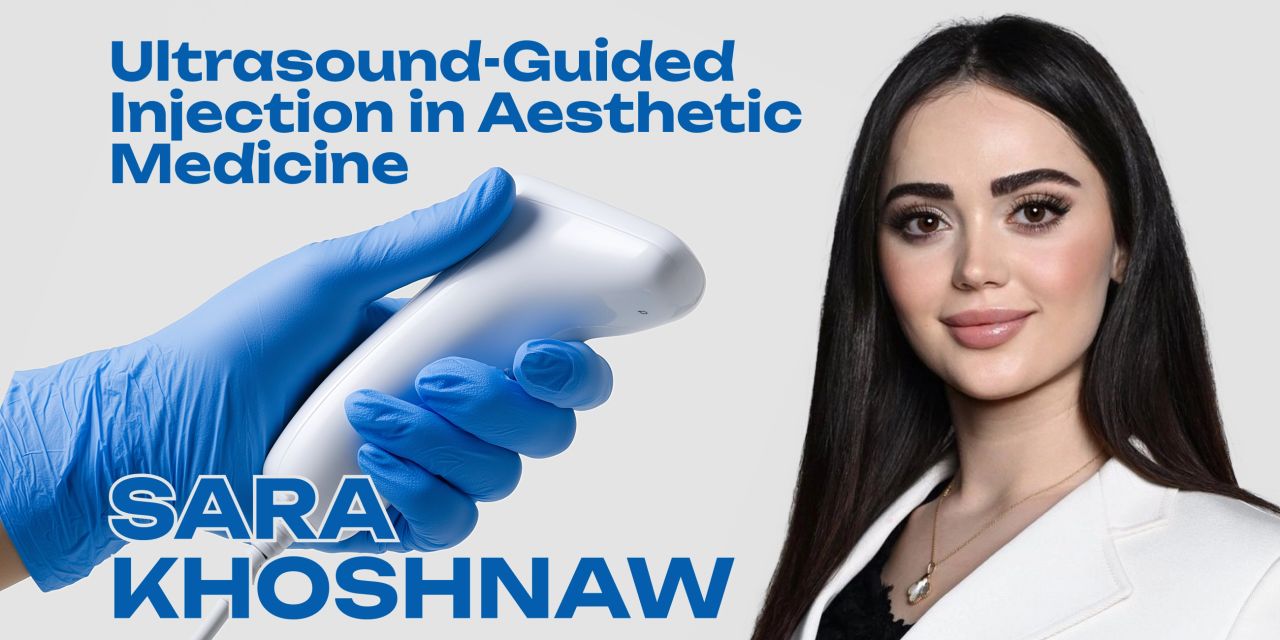
OHI-S Ultrasound-Guided Injection in Aesthetic Medicine 2 lessons 25min


$10.00
This Product is shared via google drive download link, So please share your correct Gmail id while placing the order .Please note that there are no CME points or certificate associated with this course Samples for Courses Can be found here : Free Samples Here!
EXPRESS DISCOVER PayPal
OHI-S Ultrasound-Guided Injection in Aesthetic Medicine 2 lessons 25min
Ultrasound-Guided Injections • Facial Anatomy • Dermal Fillers • Vascular Mapping • Aesthetic Medicine
2 Lessons | 25 Minutes | English
Advance beyond surface-level aesthetic injections with OHI-S Ultrasound-Guided Injection in Aesthetic Medicine 2 lessons 25min, a focused clinical course on integrating real-time ultrasound into aesthetic assessment and injection procedures.
Led by Sara Khoshnaw, an international trainer in ultrasound-guided injections, the course demonstrates how ultrasound can help clinicians visualize facial anatomy in real time, identify arteries and veins, recognize patient-specific anatomical variation, and visualize dermal fillers during aesthetic procedures.
The program connects fundamental ultrasound concepts with practical aesthetic applications across preventive assessment, diagnostic evaluation, injection guidance, filler visualization, monitoring, and therapeutic intervention.
Rather than relying exclusively on surface anatomy, the course introduces a more image-guided clinical pathway:
Visualize → Identify Anatomy → Assess Risk → Guide Injection → Monitor → Reassess
Course Details
- Course: Ultrasound-Guided Injection in Aesthetic Medicine
- Provider: OHI-S
- Instructor: Sara Khoshnaw
- Duration: 25 Minutes
- Number of Lessons: 2
- Language: English
- Primary Field: Aesthetic Medicine
- Related Fields: Dermatology, Plastic Surgery
- Primary Technology: Ultrasound
- Clinical Focus: Ultrasound-Guided Aesthetic Injections
- Major Applications: Facial Anatomy Visualization, Vascular Identification, Dermal Filler Visualization, Preventive Assessment, Diagnosis, and Therapeutic Guidance
- Primary Audience: Aesthetic Medicine Specialists, Dermatologists, Plastic Surgeons, and Other Clinical Practitioners Performing Aesthetic Procedures
OHI-S officially lists the course as 2 lessons (25min) and identifies Sara Khoshnaw as the lecturer.
Who Was the Original Program Designed For?
The course is designed for clinical practitioners seeking to integrate real-time ultrasound technology into aesthetic treatments to improve anatomical visualization and procedural predictability.
It is particularly relevant for:
- Aesthetic Medicine Specialists
- Dermatologists
- Plastic Surgeons
- Cosmetic Physicians
- Injectable Practitioners
- Clinicians Performing Dermal Filler Procedures
- Practitioners interested in ultrasound-guided aesthetic procedures
- Clinicians seeking stronger understanding of facial vascular anatomy during injections
The strongest clinical audience remains:
Aesthetic Medicine + Dermatology + Plastic Surgery
Learning Objectives
Upon completion of this course, participants should be better able to:
- Visualize real-time facial anatomy to ensure safe and accurate injections.
- Identify arteries, veins, and patient-specific anatomical variations.
- Execute ultrasound-guided dermal filler placement and perform continuous monitoring.
- Apply preventive, diagnostic, and therapeutic applications of ultrasound within aesthetic practices.
- Optimize injection techniques to achieve predictable, precise, and tailored patient outcomes.
Ultrasound in Modern Aesthetic Medicine
The first lesson introduces the role of ultrasound as an increasingly important imaging technology in aesthetic medicine.
The official OHI-S curriculum addresses:
- The role and relevance of ultrasound in modern aesthetic medicine
- Traditional aesthetic medicine approaches
- Integration of ultrasound technology into aesthetic practice
- Preventive applications
- Diagnostic applications
- Therapeutic uses
- Barriers to wider adoption
- Future potential
The lesson provides the conceptual foundation needed before progressing to ultrasound-guided injection techniques.
Traditional vs. Ultrasound-Guided Aesthetic Assessment
Traditional injectable treatments rely heavily on:
- Surface anatomy
- Clinical examination
- Palpation
- Anatomical knowledge
- External landmarks
Ultrasound adds the ability to visualize deeper structures in real time.
The course therefore introduces the transition from:
Surface-Based Assessment → Image-Assisted Anatomical Assessment
This is particularly relevant when vascular structures, previous filler placement, and anatomical variation may influence procedural planning.
Real-Time Facial Anatomy
One of the primary learning goals is to visualize facial anatomy in real time.
Ultrasound imaging can help clinicians correlate known anatomy with the actual anatomy of the individual patient.
The course emphasizes:
- Understanding facial anatomy
- Recognizing anatomical variation
- Correlating anatomy with ultrasound images
- Identifying structures relevant to aesthetic injections
This patient-specific perspective is central to the course’s approach.
Patient-Specific Anatomical Variation
Facial anatomy is not identical from one patient to another.
The official curriculum specifically includes:
Anatomical variability of the face
as an important part of ultrasound-guided injection education.
Recognizing this variability can help practitioners avoid relying exclusively on generalized anatomical maps.
The clinical pathway becomes:
General Anatomy → Individual Ultrasound Assessment → Patient-Specific Planning
Facial Vascular Anatomy
Understanding the location of vascular structures is particularly important during injectable procedures.
The course teaches clinicians to use ultrasound to help identify:
- Arteries
- Veins
- Surrounding soft tissue
- Patient-specific vascular relationships
The official lesson specifically includes distinguishing arteries and veins using ultrasound.
Arteries vs. Veins
The ability to distinguish arteries from veins helps improve interpretation of ultrasound images during aesthetic assessment.
This section connects:
Ultrasound Image → Vessel Identification → Anatomical Interpretation → Injection Planning
rather than relying solely on assumptions based on textbook vascular patterns.
Physics of Ultrasound
The second lesson includes foundational concepts involving the physics of ultrasound.
Understanding basic ultrasound principles can help clinicians interpret images more effectively and understand why structures appear differently on-screen.
This knowledge supports more meaningful use of ultrasound rather than simply operating the device mechanically.
Ultrasound Transducers
The official curriculum also covers:
Types of ultrasound transducers.
Different transducer characteristics can influence:
- Image resolution
- Imaging depth
- Field of view
- Visualization of superficial anatomy
- Suitability for aesthetic applications
The course introduces these considerations in relation to facial imaging and injectable procedures.
Correlating Anatomy with Ultrasound Imaging
A central skill in ultrasound-guided aesthetic medicine is converting an ultrasound image into a meaningful anatomical map.
The official program specifically covers:
Correlating anatomy with ultrasound imaging.
This requires the clinician to connect:
Known Anatomy + Real-Time Image + Patient Variation
to form a patient-specific understanding of the treatment area.
Ultrasound-Guided Injection Techniques
The second lesson is titled:
Ultrasound-Guided Injection Techniques in Aesthetic Medicine
and moves from foundational imaging concepts toward procedural application.
The session addresses:
- Traditional injection approaches
- Facial anatomy
- Anatomical variability
- Diagnostic ultrasound
- Therapeutic ultrasound
- Vascular identification
- Dermal filler visualization
Ultrasound-Guided Dermal Filler Placement
A major learning objective is to apply ultrasound during dermal filler placement.
The course explores the use of imaging to better understand:
- Treatment-plane anatomy
- Nearby structures
- Existing filler
- Injection location
- Patient-specific anatomy
This helps shift aesthetic injection planning toward a more visualized approach.
Dermal Filler Visualization
The official OHI-S curriculum specifically includes:
Visualization of dermal fillers with ultrasound.
This may be relevant when evaluating patients with:
- Previous filler treatments
- Uncertain filler location
- Multiple prior aesthetic procedures
- Need for anatomical reassessment
Ultrasound can therefore be used not only before treatment but also during assessment of previously treated areas.
Continuous Monitoring
The course emphasizes ultrasound not simply as a one-time diagnostic image but as a technology that can support ongoing procedural monitoring.
The educational concept is:
Assess Before Treatment → Visualize During Procedure → Monitor → Reassess
This reflects the course’s emphasis on real-time imaging.
Preventive Applications of Ultrasound
The first lesson specifically presents ultrasound as a preventive tool for early detection.
Preventive assessment can support more informed procedural planning by giving the practitioner additional information about the individual patient’s anatomy before an intervention.
Diagnostic Applications
Ultrasound also has a diagnostic role in aesthetic medicine.
The course discusses its use for evaluating anatomical structures and previously treated areas before deciding how to proceed with additional treatment.
The official curriculum explicitly includes diagnostic applications of ultrasound in aesthetic treatments.
Therapeutic Applications
The program extends beyond diagnosis to therapeutic uses of ultrasound in aesthetic procedures.
This connects imaging with intervention and enables clinicians to consider ultrasound throughout multiple stages of aesthetic care.
Preventive, Diagnostic & Therapeutic Roles
The course organizes ultrasound use across three broad areas:
Preventive
Using imaging to understand anatomy and identify relevant structures before treatment.
Diagnostic
Using ultrasound to investigate anatomy, previous filler placement, and other findings relevant to aesthetic planning.
Therapeutic
Integrating ultrasound guidance into procedural interventions.
This framework is explicitly represented in both lessons of the official OHI-S curriculum.
Improving Procedural Predictability
A key theme of the course is using better visualization to support more predictable procedures.
Rather than approaching each patient only through generalized anatomical assumptions, ultrasound allows additional information to be incorporated into treatment planning.
The overall approach is:
Visualize → Plan → Guide → Monitor → Individualize
Individualized Aesthetic Treatment
Patient-specific anatomy is one of the recurring themes of the course.
The curriculum emphasizes adapting injection planning according to what is visualized in the individual patient rather than assuming identical anatomy across all patients.
This supports the learning objective of achieving more precise and tailored patient outcomes.
Challenges in Ultrasound Adoption
The first lesson also discusses:
Challenges and barriers to wider adoption of ultrasound in aesthetic medicine.
This is an important practical topic because effective integration of ultrasound requires more than purchasing an imaging device.
Clinicians also need:
- Anatomical knowledge
- Image interpretation skills
- Familiarity with ultrasound physics
- Procedural integration
- Appropriate clinical training
Future of Ultrasound in Aesthetic Medicine
The course concludes its introductory lesson with the significance and future potential of ultrasound in aesthetic medicine.
As imaging becomes more integrated into aesthetic practice, the technology may increasingly support patient-specific anatomical assessment, procedural planning, monitoring, and complication-focused evaluation.
Instructor
Sara Khoshnaw
Sara Khoshnaw is the instructor for both lessons.
OHI-S describes her as an international trainer in ultrasound-guided injections and lists her professional activities as including speaking and lecturing at EADV, AMWC, and IMCAS, ownership of Aesthetica Private Clinic, and authorship of publications and articles in aesthetic medicine.
Her course focuses specifically on translating ultrasound imaging principles into practical applications for aesthetic injection procedures.
Major Topics Covered
- Aesthetic Medicine
- Ultrasound-Guided Injections
- Ultrasound in Aesthetic Medicine
- Facial Anatomy
- Real-Time Facial Anatomy
- Anatomical Variation
- Facial Vascular Anatomy
- Arteries
- Veins
- Vascular Identification
- Ultrasound Physics
- Ultrasound Transducers
- Ultrasound Imaging
- Anatomical Correlation
- Dermal Fillers
- Dermal Filler Visualization
- Ultrasound-Guided Filler Placement
- Injection Monitoring
- Preventive Ultrasound
- Diagnostic Ultrasound
- Therapeutic Ultrasound
- Injectable Aesthetics
- Patient-Specific Injection Planning
- Aesthetic Procedure Assessment
- Image-Guided Aesthetic Medicine
- Procedural Predictability
Who Should Take This Course?
OHI-S Ultrasound-Guided Injection in Aesthetic Medicine 2 lessons 25min is particularly relevant for:
- Aesthetic Medicine Specialists
- Dermatologists
- Plastic Surgeons
- Cosmetic Physicians
- Injectable Practitioners
- Clinicians performing dermal filler procedures
- Practitioners learning ultrasound-guided injections
- Clinicians interested in facial vascular mapping
- Professionals seeking to integrate real-time imaging into aesthetic treatment planning
The strongest clinical audience remains:
Aesthetic Medicine + Dermatology + Plastic Surgery
Why This Course Is Useful
This OHI-S course provides a concise introduction to integrating ultrasound with injectable aesthetic medicine.
The curriculum connects several important areas:
Facial Anatomy
- Real-time visualization
- Anatomical variation
- Vascular structures
Ultrasound Fundamentals
- Ultrasound physics
- Transducers
- Image interpretation
Injection Guidance
- Dermal filler placement
- Anatomical correlation
- Procedural monitoring
Clinical Applications
- Preventive assessment
- Diagnostic evaluation
- Therapeutic intervention
Individualized Treatment
- Patient-specific anatomy
- Tailored injection planning
- Improved procedural predictability
The overall clinical approach can be summarized as:
Understand Anatomy → Visualize → Identify Vessels → Plan → Guide Injection → Monitor
Product Summary
- Product: OHI-S Ultrasound-Guided Injection in Aesthetic Medicine 2 lessons 25min
- Provider: OHI-S
- Instructor: Sara Khoshnaw
- Duration: 25 Minutes
- Lessons: 2
- Language: English
- Primary Specialty: Aesthetic Medicine
- Related Specialties: Dermatology, Plastic Surgery
- Primary Technology: Ultrasound
- Major Areas: Facial Anatomy, Vascular Identification, Dermal Filler Visualization, Ultrasound-Guided Injections
- Clinical Applications: Preventive, Diagnostic, and Therapeutic Ultrasound
- Primary Audience: Aesthetic Medicine Specialists, Dermatologists, Plastic Surgeons, and Injectable Practitioners
- Educational Focus: Real-Time Ultrasound Integration in Aesthetic Injection Procedures
Short Description
OHI-S Ultrasound-Guided Injection in Aesthetic Medicine 2 lessons 25min is a focused aesthetic medicine course led by Sara Khoshnaw covering the integration of real-time ultrasound into injectable procedures.
The two-lesson program addresses facial anatomy visualization, anatomical variability, arteries and veins, ultrasound physics and transducers, dermal filler visualization, ultrasound-guided injection techniques, and preventive, diagnostic, and therapeutic applications of ultrasound in aesthetic medicine.
Topics
Lesson 1. Ultrasound in Modern Aesthetic Medicine — 13min
- Role and relevance of ultrasound in modern aesthetic medicine
- Overview of traditional aesthetic medicine approaches
- The rise and integration of ultrasound technology in aesthetics
- Ultrasound as a preventive tool for early detection
- Diagnostic applications of ultrasound in aesthetic treatments
- Therapeutic uses of ultrasound in aesthetic procedures
- Challenges and barriers to wider adoption of ultrasound
- Significance and future potential of ultrasound in aesthetic medicine
Lesson 2. Ultrasound-Guided Injection Techniques in Aesthetic Medicine — 11min
- The traditional injection approach
- Understanding facial anatomy for predictable injections
- Anatomical variability of the face
- Application of ultrasound in aesthetic injections: diagnostic and therapeutic stages
- Physics of ultrasound
- Types of ultrasound transducers
- Correlating anatomy with ultrasound imaging
- Distinguishing arteries and veins
- Visualization of dermal fillers with ultrasound
- Preventive, diagnostic, and therapeutic roles of ultrasound
Related products

Dermatology
Dermatology

Dermatology

- Anesthesiology & pain medicine
- Books
- Anesthesiology books
- Basic science
- Cardiology books
- Dermatology books
- DENTAL BOOKS
- Endocrine and Nutrition books
- Gastroenterology and Hepatology books
- Hematology / Oncology books
- INTERNAL MEDICINE BOOKS
- Nephrology / urology books
- Neurology / neurosurgery books
- Obstetrics and gynecology books
- ophthalmology / Optometry books
- ORTHOPEDIATRICS BOOKS
- Pathology books
- pediatric books
- plastic surgery books
- Psychiatric books
- PULMONARY BOOKS
- Qbank & Questions
- Radiology books
- Cardiology
- Critical Care – Emergency medicine
- DENTAL
- Dermatology
- Endocrine and Nutrition
- ENT / Otolaryngology
- Family medicine
- General Surgery
- GIT COURSES
- Hematology / Oncology
- Internal Medicine
- Medical Accounts
- Nephrology / Urology
- Neurology / neurosurgery courses
- Obstetrics & Gynecology
- Ophthalmology & Optometry
- Orthopaedic
- Pathology
- Pediatrics
- physiotherapy
- Plastic Surgery
- psychology
- Qbank & Questions
- Radiology
- Respiratory / Pulmonary
- Rheumatology
- Surgery